Wednesday, September 1, 2010

We've Lost Cedric

Cedric the Tasmanian Devil has been put down.

Cedric was at the forefront of research into the deadly Devil Facial Tumor Disease, which at last estimate was infecting 60 per cent of the wild devil population in Tasmania.

He made waves in 2007 when he was injected with a strain of DFTD and survived. He even survived a second strain with only a couple of minor tumours, which were removed.

Cedric had shown an immune response to the disease, and was otherwise healthy. It was hoped that he would lead researchers to a treatment or vaccine for the disease, but an X-ray last week showed a number of tumours in the lungs, and it was decided to euthanase the 6-year-old Cedric.

Damn.

Donations to help the Tassie Devils can be made here.

Tuesday, August 31, 2010

Cutticcini's Profile Enlargement

The wingnut Virginian Attorney General Ken Cuccinelli has had a setback in his witchhunt of Professor Michael Mann. Cuccinelli was asking for all documents from the University of Virginia concerning professor Mann's research while at UVa, to "investigate" any possible fraud committed by Professor Mann in his grants received to study climate change. A judge has ruled that Cuccinelli failed to show a sufficient “reason to believe” that UVa possessed any documents related to Mann that suggested a fraud occurred. Cuccinelli will continue harrassing UVa and Mann on the taxpayer's dime.

Cuccinelli is a climate change denier, who has also directed all state schools and colleges to remove sexual orientation references from their non-discrimination policies (in other words removed discrimination protection for gays and lesbians), and sued over the constitutionality of federal health care reform. All on the taxpayer's dime.

Cuccinelli is doing nothing more that using taxpayer money to fund a major profile enlargement, which is odd given his opposition to publicly funded healthcare.

Sunday, August 15, 2010

Evolution Among the Trilobites Part 1

The last Palaeoporn featured the growth stages of meraspid, or juvenile, Estaingia. This post will use those meraspids and a bunch of holaspids, or adults, to measure the growth patterns of Estaingia, or it’s ontogeny.

Why? Because it’s important. It allows us to plot the growth patterns and if you know the growth patterns you can used them to look at evolutionary relationships.

If you want to look at evolutionary relationships between species there is one, and only one rule.

Ya gotta know what the kids are doing.

If you don’t know what the kids are doing, you can get, as some vertebrate palaeontologists are now finding, evolutionarily embarrassed. Ontogeny can provide clues to evolutionary relationships.

Studies of ontology requires the measurement of various features to show how they change with time or size (usually the two are interchangeable – in trilobites anyway). The changes in one feature are then compared with changes in another.

At right is the head of an average trilobite showing which bits are which. This post will be concerned only with the head and what happens to it during ontogeny. Actually its even more restricted than just the head. We'll only be looking at the cranidium. That's the central area including the bulbous glabella. Why? well we need to take a bunch of measurements over a range of specimens to build up a picture of the changes that occur during ontogeny. The trouble with the librigene or free cheeks is that they are, well . . . free, and tend to be lost (as pointed out in Palaeoporn 14 and 16). The cranidium is usually left behind and so is a better subject for measurements.

At right is a cranidium of an Estaingia. The relevant measurements that we are interested in are marked. CW is cranidial width, which is literally the distance between the eyes. PGF is Pre-Glabella Field which is the area in front of the large bulbous glabella. (Incidentally the pre-glabella field only runs to the shallow trench towards the front of the cranidium. In front of the trench is the doublure. That is folded underneath the cranidium in life, along the trench, and pops up during moulting (see Palaeoporn 16). GL is the glabella length. Adding PGF and GL gives us a value for the length of the craniduum. POG is Pre-Orbital Glabella which is the bit of the glabella that lies in front of a line drawn between the front tip of the eyes. AF-EL is the distance between the end of the Axial Furrow (the trench behind the glabella and the back tip of the Eye Lobe.

Lets show some measurements - the length of the central portion of the trilobite head, the cranidium, compared with the width of the cranidium, and the length of the glabella, for each specimen, as seen below
Here the measurements fall on a straight line. This is called isometric growth, where the ratio of the two features does not vary with growth. In example A, the ratio of the length of the cranidium to the width of the cranidium does not change with growth. In example B the ratio of the cradidial length to glabella length does not change. So during isotmetric growth there is no change in the shape. (What? Yes, OK, example A is not strictly isometric, since isometric growth always passes through the origin and a line drawn through the plot in A doesn’t. Just. But trust me, compared with what’s coming up next it’s pretty much isometric.)

Isometric growth can be very useful in confirming that specimens belong to a particular species. In the examples above, the red dots represent meraspid Estaingia and the blue dots holaspid, or adult forms. The fact that they all line up on a straight line is supporting evidence that the meraspids do belong to Estaingia. This is important because species are defined by a “type” specimen which is normally an adult, and so it can be quite difficult to place meraspids in the correct species if the growth patterns vary considerably from the adult.

Oh yes, the black dots at the far end of both graph trends represents a holaspid Estaingia from a different location and a slightly younger deposit, at Cymbric Vale in New South Wales (NSW Estaingia). Thought to be the same species as E. bilobata discussed here from the Emu Bay Shale, but also though not to be for a couple of reasons. It'll feature later but see how it plots along the same trend as the other specimens? good evidence that it is E. bilobata, but I'm getting ahead of myself.

Frankly, however, isometric growth while it has it’s place, is a bit boring. Meraspid 1 looks pretty much like the adult form. (no problem in putting the meraspids in the correct species there then.)

Just as well then that not all growth is isometric. In fact most growth patterns are not isometric, but anisometric. Anisometric growth, as you may suspect, is where the ratio does not remain constant, but changes during growth, and so shape changes during growth. Which means the meraspid can look distinctly different from the holaspid.

Holy rotating vectors Batman!

Now it gets interesting. There is only so much you can do with straight lines. Everything is better with curves! Anisometric growth changes are not random. They don’t occur because the merasid didn’t like the look of itself in the mirror. The represent hereditary traits. In the two graphs above, A shows Cranidial Width against Axial Furrow to posterior of Eye Lobe (as a percentage of cranidial lenght) (CW/AF-EL) and B shows Cranidial Length against Pre-Glabella Field (as a percentage of cranidial length) (PGF/GL+PGF).

Here the difference in growth patterns between the merasipd and holaspids is marked. This is telling us important information about how the final look of the holaspid form is achieved, and what pattern of growth occurred.

In graph A the feature being measured is the curvature of the eye. In early meraspids, the eye curvature is quite shallow and so the lower end of the eye is a significant distance from the Axial Furrow. As the meraspid grows, the eye curvature becomes more pronounced and the distance between the lower end of the eye and the axial furrow decreases. At the end of the meraspid stage, the distance (as a percentage of cranidial length) is around 0.25. In the holaspid stage this trend ceases, the eye reaches its maximum curvature, and doesn't change again, so the value remains at around 0.25. The NSW Estaingia plots along this holaspid trend albeit at a larger size that those at Emu Bay, but again, it is good evidence that NSW Estaingia is Estaingia bilobata.

In Graph B the preglabella field as a percentage of total cranidial length is plotted against cranidial length. Here the trends are different. In the meraspids, the preglabella field increases with growth until the Holaspid stage is reached. Then the trend is reversed and the preglabella field begins to reduce in size with growth, albeit at a slower rate than the meraspid rate. The NSW Estaingia again plot on the holaspid trend for Estaingia bilobata providing more evidence that the NSW Estaingia is E. bilobata.

One last graph which shows the preorbital glabella or POG (the portion of the glabella in front of the eyes).

Note that this is a mirror of the pregalbella field plot, and provides an explanation for the pregalbella field changes. The preorbital glabella in the meraspids decreases in size relative to cranidial length. In other words, as the cranidium increases in size, the preorbital glabella is increasing at a slower rate. This means that the area in front of it has to increase in size. So the increase in the pregalbella field we saw in the previous chart is a result of the preoccular glabella not keeping up during meraspid growth. However, in the holaspid stage, this trend is reversed and the preoccular glabella starts to grow at a more rapid pace than the cranidium generally, and begins to represent more and more of the total cranidial length. Thus the preglabella field begins to decrease as a proportion of the total cranidial length. Once again the NSW Estaingia plot among the Estaingia bilobata and along the trend. We can say with some confidence then that NSW Estaingia is a larger version of Estaingia bilobata.

Next time how these growth plots can provide information on evolutionary relationships.

Saturday, August 7, 2010

2.1 Ga Multicellular Colonial Organisms - Update 2


In posts here and here I discussed why I thought the 2.1 Ga structures from Gabon, figured in the Nature paper, were actually microbial mats and not examples of multicellular colonial organisms.

I now have some further info which strengthens my view. I'd like to thank Dr Diana Cuadrado of the Instituto Argentino de Oceanografía who very kindly sent me some more images of microbial mats.

This mainly revolves around the claim in the paper that microbial mats . .
. . often leave characteristic in carbonate ad siliciclatic rocks. Such structures, however, including those formed in shales and mudstones, do not resemble the Gabon fossils". (p. 103)
As I've discussed previously, I disagree.

At the top of this post is an example of a modern microbial mat (top image), compared with a photo of the 2.1 Ga structures (bottom image). The black box in the top image represents the area of the lower image at the same scale (The bar scale is 10 cms in the top image and 1 cm in the bottom image). This reinforces just how small these 2.1 Ga structures are.

Here then, is more evidence that microbial mats can produce the structures seen in the 2.1 Ga specimens.


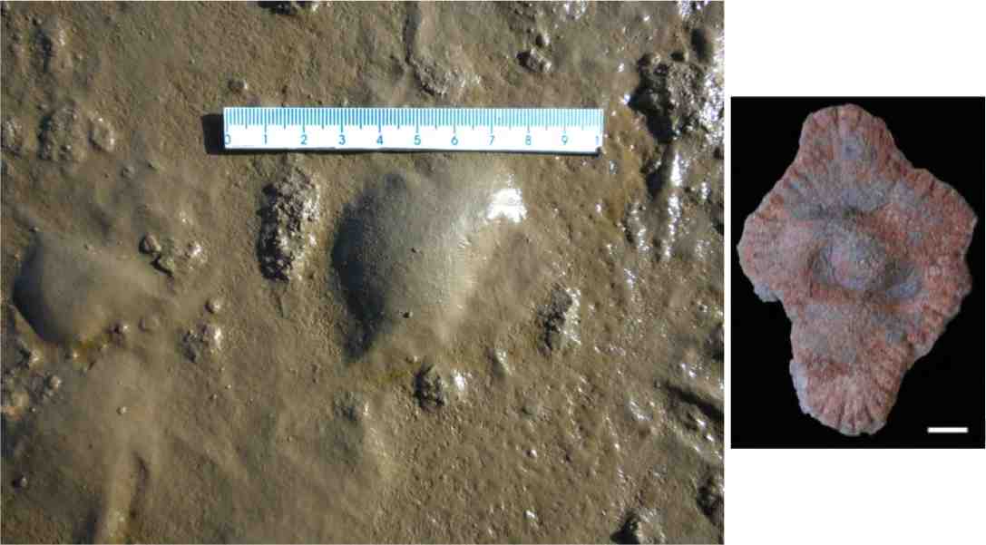
Here is a close up of a large gas bubble in a modern microbial mat (it was taken last week - you can't get much more modern than that!). As you can see by the comparison with one of the 2.1 Ga specimens at the same scale, gas bubbles in microbial mats are on the same scale as the 2.1 Ga specimens.

For me this is the clincher (if a clincher were needed)


The top photo is of a modern, ruptured gas bubble, the bottom is of a 2.1 Ga specimen. See how the modern microbial mat is flexible enough to fold and stay intact even when torn. But, see the folding to the top and right of the hole. It's almost an exact match for one of the 2.1 Ga specimens figured, to scale, below. No that's not quite right . . . it's an exact match!

Not only that but there is a small fringe beyond where the folds end in the modern example, similar to that seen in the 2.1 Ga specimen.

To say something like "Ta Da" at this point would be churlish, juvenile and unprofessional, . . . so

TA DA!

But wait, there's more.


This is a photo of a mature microbial mate with a couple of overturned pieces. Not the cracking in the larger piece around the margin. his may be the cause of the "radial fabric" of the 2.1 GA strucutures. The smaller piece has even more marked cracking and looks similar to this 2.1 Ga specimen below.


This latest evidence strengthens the argument that the 2.1 Ga structures are pyritised microbial mats and not multicellular colonial organisms.

Thursday, July 29, 2010

Palaeoporn 17

Meraspid

Drumming

You never forget your first. The time. The place. The moment.

The windswept coast. The bed that refused to cooperate. A cleavage that simply wouldn’t quit. But then suddenly, everything is revealed. The curves! So different. Stood out in the crowd despite being petite. I couldn’t fail to see, even at a distance of 4 feet.

No, you never forget your first. First meraspid that is.

This fellow is a personal favourite of mine. It’s a meraspid (or juvenile) Estaingia bilobata It was the first meraspid found at from the Emu Bay Shale. It’s a stage 10 meraspid. Thanks to it, and other merasipds found in the Emu Bay Shale, we can work out the ontogeny or growth pattern of Estaingia, and as an added bonus allows us to work out some evolutionary relationships (but that’s for another post).

This Palaeoporn is all about ontogeny amongst the trilobites, more specifically Estaingia.

One of the great things about trilobites is that, to grow, they have to moult the hard exoskeleton. This means that one trilobite can leave behind a complete growth history in moults. Since this is unlikely to happen for one, many trilobites of the same species leaving moults behind allows us to build up a picture of the growth pattern in a way that we simply can't do for moult-challenged organisms.

Meraspids are what trilobite larvae grow up to be before they grow up to be adult trilobites. Trilobite larvae, or protaspids look like just a head. Suddenly things get serious and the protaspid gets a tail. Or at least the beginnings of one. A furrow appears towards the back of the protaspid, which will separate the tail or pygidium from the head or cephalon. Once the furrow is complete, the trilobite is considered to have moved into the meraspid stage - meraspid 0. 0 because it has no thorax segments yet. Just a head and a tail.

Once in the meraspid stage, the trilobite begins to add segments to the thorax, usually one per moult, but in some species this can be more. Each meraspid degree or stage is represented by the addition of another thorasic segment and is numbered according to the number of segments.

The specimen at right (Fig. A) is a Meraspid 1, the smallest meraspid so far found at Emu Bay.

The most striking thing (apart from it's size - the white scale bar represents 1 millimetre! Hence it's a bit blurry) is the spines. In very small forms spines become important. If meraspid 1 was swimming amongst the plankton, the spines would help with buoyancy, helping keep the meraspid afloat. If the meraspid was on the sea floor, then the spines would help keep the meraspid from sinking into the mud. We don't know if Estaingia meraspids were planktonic or scurried around on the sea floor. The spines may also provide some defense, but the get smaller as the meraspid grows so it doesn't appear that defense was a primary function. In meraspid 1 the one segment has a pair of large spines, as well as a pair of backward pointing spines on either side of the head. Meraspid 2 would have had two segments each with a pair of long spines.


As the meraspid grows, segments are added from the front of the tail. We can see this in meraspid 7 at right (Fig. B, scale bar 1 mm). This has 7 segments, but the two original segments with the spines can be seen at the front of the thorax, with the non-spiney segments, which have been added later, positioned behind these two. If the thorasic segments were added from the back of the head, then the first two segments with spines would be at the back of the thorax next to the tail. The large spines on the head are also still very visible.


In Meraspid 10 at right (Fig. C, scale bar 1 mm) things are starting to change. The first thing to notice is that the large spine on segment 1 is almost gone. There is a much reduced spine there. Also the spines have become more curved. They are still long though - still the length of the meraspid. The head is very well preserved and provides a wealth of information on the way the various elements of the head change with growth. The rounded shape and the spines are very distinctive. Nothing else in the fauna is like that. So, even though it's only a few millimetres across, it immediately caught my eye.


Finally we have meraspid 12 (Fig. D, scale bar 1 mm). We are almost at the Holaspid stage or mature stage, of 13 segments, and the meraspid is now similar to the final adult shape. Note that the second thorasic spines are much reduced, and the spines on the head are also beginning to reduce in length. The thorasic spines will completely disappear in the adult, and the spines on the head will be reduced, but still present.


And this at right is a fully mature Estaingia. You can see that the thorasic spines have gone, and the spines on the head are much reduced.


Below is a diagrammatic representation of the ontogeny of Estaingia featuring the four meraspids figured above.


It gives you a better idea about what changes are occurring as the trilobite matures. Features to look out for are the relative width and length of the head and the glabella or central bulb region of the head. This is important because these relative values can be plotted and, as they remain at a constant ratio can help confirm the identification of the meraspids with the holaspid form. Like so:

This is a plot of the length of the longest part of the head (cranidium) against the width from eye to eye, and the longest part of the head against the longest part of the glabella - the central bulb area of the head.

As you can see the meraspid measurements (in red) line up along the same trend line as the adults (in blue) giving us a high degree of confidence that the meraspids are of Estaingia. Also notice that the measurements are clustered - i.e. not evenly distributed. This represents the moulting habit of the trilobites. As growth can only occur once the trilobite has moulted the pattern of growth is stepped. Unlike a constantly growing form, where the plot would be more even.

Using measurements of growth patterns can be very useful because with some trilobites, varying the time spent in particular growth patterns as can dramatically alter the end result - or what we call evolution. But that's for another post.

Monday, July 26, 2010

Informed about Introns

Stephen Matheson has a series of posts about introns and their misuse by the Disco 'Tute.

Genes are transcribed into RNA for shipping out of the nucleus to make proteins. Introns are areas of the RNA that have been transcribed from the gene but are then removed from the RNA template prior to it moving out of the nucleus.

It all about the junk - or not-junk.

Go read Steve's posts Part I, Part II, and Part III, and get informed.

Thursday, July 22, 2010

Palaeoporn 16

Extreme Ecdysis


When you're broken, in a million little pieces . . .

You may want to check out Palaeoporn 14 and 15 before reading this as Palaeoporn 16 follows on from them. That's ok. We'll just talk amongst ourselves until you get back.

So . . . where are you from? . . . Fort Saint John, Canada? Really? Good ol' Highway 97. . . . umm, come here often? . . .

Oh, ok you're back. Right. First thing, you need to see this video of a giant spider crab moulting (hat tip PZ)


Once you've seen it and read PZ's piece on moulting, or ecdysis. The linked video of a spider crab moulting in PZ's piece is the one featured here. Very instructive, if not a little creepy. But remember that trilobites - being proper arthropods - exited the old body exoskeleton old school i.e. head first, not arse first like a common chelicerate.

Ok, now we have an idea of what ecdysis is, this Palaeoporn is an example of what happens when things go wrong . . . very wrong.

The photo at the top is of the trilobite Redlichia takooensis, or rather what's left of one after a particularly difficult mould. Just to remind you, at right is what a Redlichia takooensis should look like. R takooensis is a Lower Cambrian trilobite with a 15 segment thorax and small pygidium or tail. A major characteristic of R takooensis are the large spines, on either side of the head, at the back of the head in the centre, and two large spines on the axial rings of segments 6 and 11 (counting back from the head) (What? Axial ring. The middle part of the thorax. What? Thorax. The middle part of the animal between the head and the back end (pygidium). What? Yes the pygidium is very small, it's a micropygous trilobite. What? No. The division head-thorax-pygidium is not how the trilobite got it's name. The "three lobes" refers to the three zones of the thorax, the plura on the left, the axial ring, and the plura on the right).

There are probably spines on each axial ring, but 6 and 11 have the biggest. The spine coming off the rear of the trilobite, seen in the photo (above right), is from segment 11 - so the spines were pretty long.

To give an idea of what R. takooensis looked like I'm going to cheat an use another trilobite to approximate Redlichia.

Left side view of Paradoxides masquerading as Redlichia (it's got three extra segments - no, don't count them!). Thick line represents facial suture. (adapted from Whittington 1990)

Notice the spines. These will be very important in what happens in extreme ecdysis. In life, the spines would have not sat flat along the thorax, but would have been mobile and could be moved in an arc, from flat along the body (as above and in complete trilobite photo above right), through some acute angle (as seen in the ParaRedlichia diagram above).

What you don't see is the series on notches on the back margin of the first 5 segments of the thorax. These would have acted to lock the first 6 segments in place making this portion of the thorax act as a rigid body. That's also important.

What's also important is that the trilobite could bend. It could flex into an arc - though not into a ball like later trilobites (an important feature for Anomalocaris as discussed previously). Also the maximum flexure occured around segment 5.

OK, ecdysis in Redlichia. Normally Redlichia would have operated in a similar way to Paradoxides shown below.

Maneuvers during exuviation of Paradoxides 2. Left side view, thick line on head represents the suture line. 3. Flexure of body. 4. Front view showing open sutures and animal beginning to emerge (stippled). 5. Side view of animal beginning to emerge. (Whittington 1990).

The trilobite arched upward, planted the front of the head in the sediment and pushed using the back end as a brace. This put pressure on the head and the sutures around it. These part, with the cheeks on either side parting from the central area of the head, and the front part of the head separating and lifting upwards. This allowed the soft new body to emerge through the front of the head and 'head' off, leaving the cast of exoskeleton behind.

There is one difference (alright two). The spines. These could have been brought into play if the old exoskeleton didn't play ball, as shown below.


Here, the rear spine could have been used as a lever to apply more pressure on the head sutures, forcing them to separate and allow the trilobite to exit. How do we know this? well, a) it seems like a reasonable thing to do, and b) we have the specimen figured as Palaeoporn 16 (and others).

Let's remind ourselves of the specimen, 'cos its been a while.


What we have here is one disarticulated trilobite. There is a split in the thorax between segments 4 and 5, and another between segments 10 and 11. The thorax has been split into three pieces, comprising segments 1-4, 5-10, and 11-15 (plus pygidium).

Which brings us to a very important question. Where's the head?

The left free cheek is there, roughly where it should be at the front end, but it's inverted. It's inside out. Flipped over. The nice curved margin you can see is actually where it joined the eye. But the rest of the head?

Well, most is missing, but see the white and orange line running away from the free cheek and pointing to the 2 o'clock position? That's what's left of the head. It's the lower part of the head, basically a thin band of exoskeleton (called the doublure) which runs around the underside of the head and attached to both free cheeks and the front of the central portion of the head (the doublure is show in black at right). That central top portion of the head is gone, but the doublure is rammed into the sediment and twisted.

To give you some idea, here is a somewhat stylised diagram to illustrate.


What we have is one very sad and sorry trilobite. But, knowing what we know of ecdysis in Redlichia we can piece (Ha!) together what happened.

Knowing that the first 6 segments could lock together, and and the spine on segment 11 was used as a pivot point, simple mechanics tells us the the maximum forces exerted at or around segment 5, (where the maximum curve would be - the locking of segments 1-6 would re-enforce this), and around segment 11.

So, the trilobite arched up, rammed the front of the head into the sediment and braced with the spine on segment 11. Nothing happened.

Eventually the arching and the bracing built up so much pressure that something had to give. In very quick succession, the exoskeleton behind the head went (we know this can happen from Palaeoporn 15), as did the sutures on the head. But also the pressure acting on segment 5 became too much and the exoskeleton ruptured between segments 4 and 5. Also to pressure around segment 11, caused by bracing against the spine, causes the exoskeleton to rupture between segments 10 and 11.

The trilobite exited the broken exoskeleton, probably taking the middle portion of the head with it. That left the free cheeks separated from the central head exoskeleton, but still attached to the doublure, which was buried in the sediment. The left free cheek, still attached on its outer side, rotated outward to lie inside out, while the other is buried.

Tough moult!

But there's more. Here's another example. Similar story with similar results (sorry it's in black and white. I could say it's in black and white for the ambiance, but I just don't have a colour photo of it).


This time the thorax exoskeleton gave way behind segment 6 and behind segment 11. And this time we have the head. And both free cheeks, but they are inverted an displaced slightly. Also the doublure is present. it's the thick band right at the front of the head.

This is a classic example of a broken, incomplete, trilobite providing more information than a perfect specimen would. They provide information on trilobite activity and mechanical information on the exoskeleton.

They also provide a whole new definition of the term "growing pains"


Whittington, H.B (1990) Articulation and Exuviation in Cambrian Trilobites. Phil. Trans. R. Soc. Lond. B. Vol. 329 no. 1252. pp:27-46 doi:10.1098/rstb.1990.0147

Monday, July 19, 2010

Never Listen to the Weather Forecast

Never listen to the weather forecast.

The forecast of an impending heavy thunderstorm.

An impending heavy thunderstorm that threatened to overflow your blocked gutters.

The gutters you decided to clean with the help of a ladder.

The ladder you don't secure well enough so that you fell off.

A fall of about 15 feet to the ground.

The ground from which your housemate had to drive you to hospital.

The hospital where you stayed 10 days.

Ten days in which they scraped together what's left of your elbow.

The elbow that needed a metal implant to function and so looks like this.


(A metal implant that doesn't set off the metal detectors at airport so that you can't even look at the security people and nod and say 'yeah, that's me')

A metal implant which required 8 months of physio come rain or shine.

The rain that reminded me of that forecast heavy thunderstorm that NEVER CAME.

So yeah, never listen to the weather forecast.

On the up side, I can now get all my images (x-rays, CT) on a cd which I can keep and upload to the internet!

Tuesday, July 13, 2010

2.1 Ga Multicellular Colonial Organisms - Umm, Not (update)

I previously discussed why I don't think the 2.1 Ga structures found in Gabon are multicellular colonial fossils.

Having looked at the Nature paper some more, another thing springs to mind.

Scale.

Here are some of the structures as figured in the paper.


Note the scale bars against each example. Those scale bars represent 5 mm! That's right millimetres!

These things are small. Especially the central area that contains the folding.

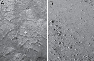
Now, compare that with the examples of bubbles in microbial mats

Photo from Cuadrado and Pazini (2007)

There is no scale, but some of the bubbles must be at least the size of the central folded area of the specimens figured in the Nature paper. This means we have structures documented today in microbial mats that equate in size to the central area of the structures from Gabon.

Now, what would happen to a gas or fluid filled bubble if it were covered with sediment and the gas/fluid escaped? It would deflate. Not deflate flat, as the microbial structure would be too thick. Maybe it would fold on itself just like the central structure in the specimen in row two above? Especially if there was an encouraging push from overlying sediment.

I'm even more convinced that the Gabon structures are microbial mats.

UPDATE 2

Diana G. Cuadrado and Natalia V. Pizani. (2007) Identification of microbially induced sedimentary structures over a tidal flat. Latin American Journal of Sedimentology and Basin Analysis. v.14 n.2 La Plata ago./dic. 2007

Monday, July 5, 2010

2.1 Ga Multicellular Colonial Organisms - Umm, Not


ResearchBlogging.org

2.1 GA structures on black shale bedding surface. Scale bar 1 cm. (El Albani et al. 2010)


The latest edition of Nature has a paper claiming 2.1 billion year (Ga) old multicellular colonial organisms from Gabon. This is a remarkable claim, since the oldest definitive large multicellular organisms so far found in the fossil record are from less than 600 million years ago (It is still not certain if the 2 Ga Grypania spiralis is a eukaryote or large bacterial colony). So remarkable is the claim in fact, that it made the front cover of Nature.

I think they are wrong.

Lets be more specific here. There are two claims - that the structures are 2.1 Ga old, and that they represent large multicellular colonial organisms.

First the date. That appears well constrained. Chris Rowan has good coverage of the dating at Highly Allochthonous, so I have no argument against the age.

I don’t think they are multicellular colonial organisms. I think they are pyritised remains of microbial mats, or structures associated with microbial mats.

There’s been ample coverage of the paper, but I want to concentrate on the reasons why the authors think that the structures represent organised colonial organisms.

The structures are found in unmetamorphosed black shales. Over 250 specimens have been recovered. They are pyritised and embedded within the sediment. They range from 7-120mm in length, 5-70 mm in width, and 1-10 mm thick. They can occur in densities of up to 40 specimens per square metre, at random orientations, but all are horizontal to bedding. All are wrinkled to some degree, with some having large central pyrite nodules. Some show significant folding. All show radial cracks. The authors used micro-computed tomography (Micro-CT) to produce three-dimensional images of the structures. They also undertook carbon and sulphur isotope analyses of the host rock and the structures.

On the following lines of evidence the authors decided that the structures represented multicellular colonial organisms:

  1. The structures shown under Mirco-CT are not the same as produced by microbial mats.

  2. They are unaware of any inorganic process that could produce the folding patters seen in the structures.

  3. The folding suggests an originally cohesive flexible sheet.

  4. They are unaware of any inorganic process that could produce the radial fabric seen in the structures.

  5. The radial fabric represents peripheral accretion of flexible organic matter.

  6. The 12C/13C carbon isotope ratio in the host sediment is different to that in the structures suggesting the structures represent distinct organisms.

  7. Steranes have been found in the shales, a compound associated with eukaryotes.

  8. A lack of support for any other inorganic origin.
I think pyritisation of microbial mats is a better explanation. Here’s why.

I’d like to take 1, 2, and 3, together because they all relate to the physical properties of microbial mats.

First let's take a look at what we're talking about.

Micro-CT-based reconstructions and virtual sections of four specimens from the FB2 member of the Francevillian Group. First Column - original. Second column - volume rendering in semi-transparency. Column 3 - Transverse two-dimensional specimen. Column 4 - longitudinal section running close to estimated centre of specimen. Scale 5 mm. (El Albani et al. 2010)

The structures are pretty much all pyrite. The absence of pyrite is marked by the radial cracks. The bright central area in rows 3 and 4 represent a central pyrite nodule. This is not present in all specimens. This is important, as the large thickness values for specimens are all associated with this central nodule. Away from the nodule, or when a nodule is not present (e.g. row 2 above), the structure is a thin film of pyrite around 1-2 mm thick.

This is really important. All the folding is just that, folding. The apparent holes (black areas) in the images above do not represent complex structure within the specimen. The image is a slice through the specimen at a certain level. Where the pyrite film is folded below the level being imaged (downward folds), or completely above it (upward fold), you get a black space. The images appear to show complex structure, but it is simply a line drawn across an undulating surface. There is no internal structure here other than the radial fabric.

Further, and critically, the authors have concluded that the thin pyrite film and the central nodule represent different periods of pyrite formation. This is supported by sulphur isotope data, and I am happy to accept their conclusion.

This means that the large nodules have little to do with the original structure of the specimens, and, in fact they appear to occupy a central cavity in the specimen when they are present. An example of an actual nodule is below.

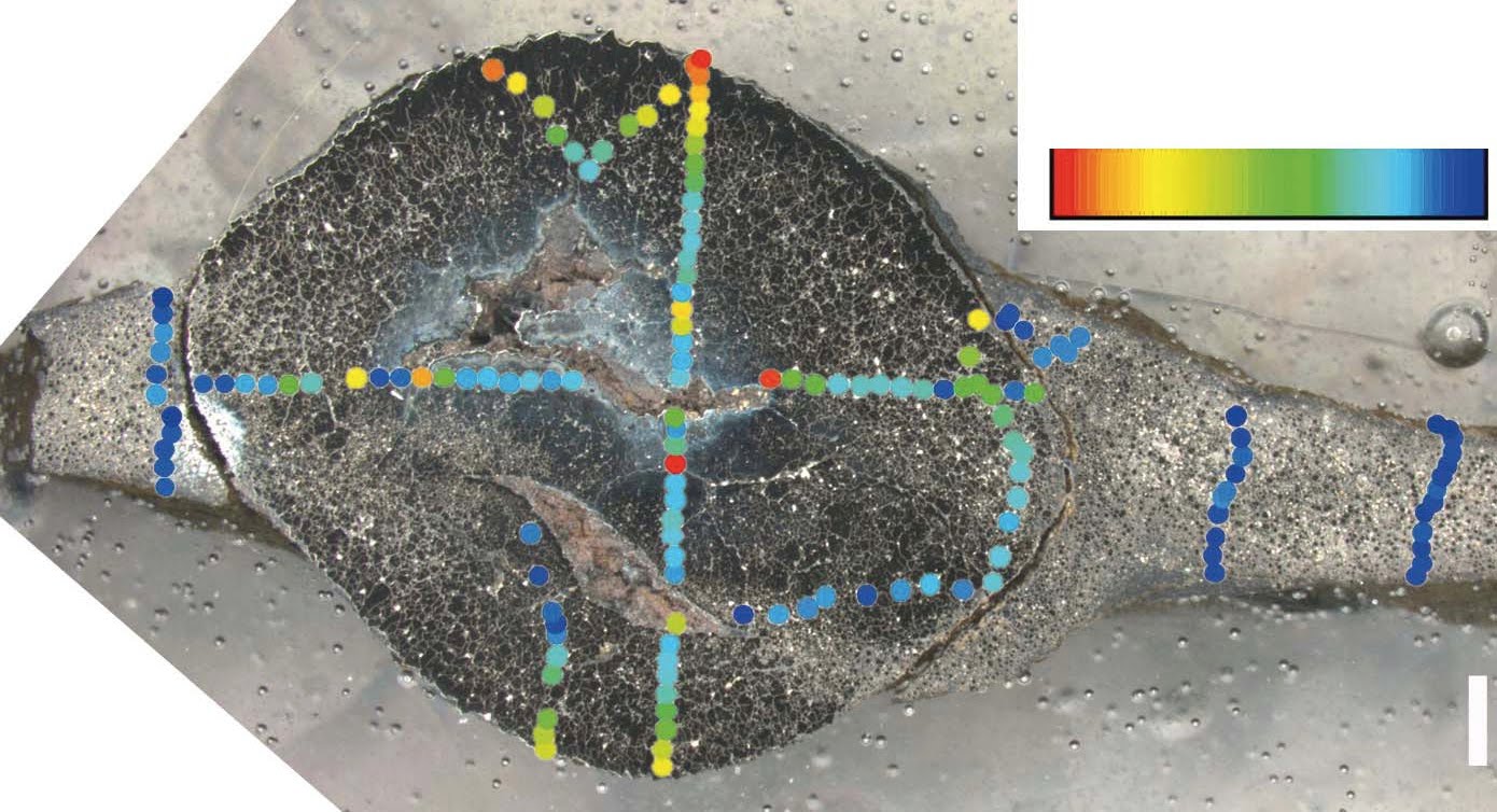
Section through a specimen showing central pyrite nodule surrounded on both sides by sheet material. Scale bar 1 mm. (El Albani et al. 2010)

In this specimen you can see that the nodule and the thin film on either side of it is almost all pyrite. The gaps in the center of the nodule are unexplained, but probably host sediment. Ignore the pretty colours. that refers to sulphur isotope analysis which I'm not questioning.

So, if we ignore the central nodules for the present, we are left with an original structure that is a thin film approx. 1-2 mm thick which has been thrown into folds in some specimens (e.g. row 2 above) but not in others (e.g. row 1 above).

The authors claim that the folding and the radial cracks cannot be accounted for by inorganic processes. I think they can.

Look! over there on the right! Is a 2.1 Ga colonial organism with a radial fabric? Is it a Proterozoic jellyfish? No, it's . . well, I'll explain later. But compare this example with rows 2, 3 and especially 4 of the Micro-CT images above. Notice that it has a central zone which is distinct from the periphery. Notice there is a faint dark band separating the central core from the periphery. Notice also the distinctive radial 'cracks' that spread out from the core Central mass? Check, radial cracks? Check. This would appear to be a good match to the 2.1 Ga structures.

But if you are not convinced, here's another variety of the same structure. this time we'll compare it directly with row 2 above as this has the best fold structure presented.


Can you see the fold pattern at the centre of the lower photos? It's quite similar to the fold pattern in the Micro-CT image. These images are of water escape structures in Ediacaran sandstones. The difference in colour you can see is due to the sandstone being deposited wet and dirty - i.e. waterlogged and with a significant amount of silt and clay (the red stuff). Escaping water carries the red silts and clays away with it, leaving the white sand behind.

Three things to note. They can produce radial structure, they can produce fold-like structures, and they are pretty similar in size even though they are from different sandstones.

Now, for the record, I don't think that the radial structures in the 2.1 Ga structures are water escape features (I've another idea for them), I'm just showing that such structures can be formed inorganically. Although I am suggesting that water escape could be an explanation for the folding we see in the row 2 specimen.

The authors discount water escape as an explanation because the underlying sediment shows no disruption such as that shown in my Ediacaran example viewed from the side. Plus, shales are not known for their permeability (ability to allow water to flow through it). But we are dealing here with microbial mats. Mats can act as a barrier to water or gas flowing below them. In such instances, rapid loading by waning storm sediments could cause water or gas trapped under the microbial mats to tear through the mat, creating some neat folding patterns and not disturb the underlying sediment too much.

Actually I'd better show you some mats so that you know what I'm talking about. Here's one.

A juicy, yummy microbial mat, full of cyanobacterial goodness from Yellowstone Park. Photo Carnegie Institution

(And yes I do consider Yellowstone to be a good analogue for the 2.1 Ga Proterozoic, because while the hydrothermal pools are aggressive to microbial mat grazers, so was the 2.1 Ga Proterozoic, because, well, there weren't any.)

Do you think the microbial mat above looks like what the authors have imaged? I do.

And just so we're clear on how flexible microbial mats can be, here's another photo.

Photo from Cuadrado and Pazini (2007)

Here are some more Microbial mats.

Photo from Cuadrado and Pazini (2007)

Photo A shows microbial mats draped (folded) over cracks. Photo B shows gas bubbles trapped under a microbial mat. What? you thought I just made that gas stuff up?!

But this is the clincher for me.

Microbial mats at West Chaplin Lake (Bowman and Sachs 2008)

There are a number of things to notice here. Firstly the mats are not one continuous sheet, but here are two discrete mats about the same dimensions as the 2.1 Ga structures. Also note the lower mat has been torn to produce a clean, sharp, high angled edge. This shows that these things can be torn without losing structure, so simple folding should be no problem. Speaking of folding, check out the left margin of the upper mat. See the fold? Remind you of anything? This maybe?


So microbial mats can fold and wrinkle without tearing. I agree with the authors that the structures represent originally cohesive flexible sheets, but microbial mats not colonial multicellular organisms.

There are other methods to fold or wrinkle microbial mats. Storms could rip up mats or partially dislodge them for example, and the authors interpret the environment as deltaic, influenced by storms. Elephant skin textures on bedding plains have been interpreted as being caused by wrinkling and folding microbial mats.

I hope this has shown that the structures seen in the 2.1 Ga specimens, the folding and wrinkling, can be produced by inorganic means on biological sheets without invoking a new class of organism.

OK, on to numbers 4 and 5, the radial fabric.

This is one of the main arguments for the structures being a colonial multicellular colonial organism and it certainly is striking. but, as I showed above, radial fabrics can be caused by other things. inorganic things.

This irregular radial fabric, along with the the fact that it is commonly deflected to meet the edge of the structures is taken as evidence that the structures grew by peripheral accretion of flexible organic matter.

However, there are a few specimens where this radial fabric appears to go right through the structure.
Scale bar 1 cm

In one example, the fabric appear to actually cut through the central nodule

Scale bars 5 mm


How could a growth-related fabric cut through a nodule that wasn't there until late in the diagenetic process?

I think it's down to water again.

Lets assume that the mats/colonial organisms have gone through initial diagenesis and have been converted to pyrite. As diagenesis proceeds and load on the sediment column increase, the mudstones start to compact. Muds can contain 60% water which means that they have a long way to compact. Problem. Pyrite sheets can't compact.

I think that as compaction progressed and the pressure increased, the thin outer margin split in a regular pattern. As the process continued, the cracks spread further into the body of the specimens and sediment would be forced into the cracks. As we have an example of crack through the central nodule, it may be that this cracking occurred late in the diagenetic process, when compaction was reaching it greatest extent.

So rather than be evidence of peripheral growth, the fabric may be diagenetic.

Number 6 and 7 are linked, 6 is the carbon isotope ratio, and 7 the presence of steranes.

The authors found a difference in the organic carbon isotopic composition between the host rock and the structures. This, they say, shows that the structures were distinct organisms.

Without going into too much detail, carbon is present in two main isotopes 12C and 13C with one extra neutron in the nucleus. Organisms preferentially take up 12C in different amounts depending on their metabolic pathways, causing a shift in the 12C/13C ratio. Measuring this difference or delta 13C can help identify the origin of the organic mater.

The host rock has a delta 13C value of -27 while the structures had a value of -32. Now -27 is roughly an average value for eukaryotes, and so would be expected to represent the fallout from the water column into the sediment of dead eukaryote plankton. A value of -32 is more like bacterial signature. This would tend to support my suggestion that these structures are pyritised microbial mat fragments.

Number 7 is the observation that steranes have been found in the shales (note the shales not the structures). Steranes are compounds associated with eukaryotes. But if the explanation for the delta 13C values is correct, the steranes would also be produced from the eukaryote plankton and can't be used to support the suggestion that the structures are eukaryotes.

Finally number 8, a lack of support for any other inorganic origin.

I think I've put up enough suggestions for inorganic input into the formation of these structures. but here is a rough explanation:

- Microbial mats grow on delta front muds - probably as individual round patches, possibly connected by thin connections to other mats.

- Storm activity rips up or distorts the mats into folds and waning storm currents deposit a layer of sediment over them.

- Pore fluids equilibriate and the mats are surrounded by sulphide dominated pore fluids.

- Pyrite replaces the mats.

- As the basin fills and the sediment column increases, the muds compact. The distortion and pressure casuse the pyrite structures to deform slightly and crack around the margins.

- As pressure and compaction increase the cracks propagate and second pyrite phase occurs in the central area of some structures. It may be that the cracks allow pore fluid and organic-rich sediment into the centre of the structure allowing the second pyrite phase to occur.

I'd like to commend the authors for taking a multidisciplined approach to this work. It's an approach which I believe should be done more often, and will be more widespread in the future.

I'd also like to commend them for their use of new techniques and I hope they will continue to use them to explore more of the geological record.

Sorry that this post has been a bit of a smorgasbord, but my aim is to present a number of possible scenarios for the formation of these structures that do not require that they be large, multicellular, colonial organisms. There are other, unfortunately more mundane, explanations.


UPDATE 1

UPDATE 2


Albani, A., Bengtson, S., Canfield, D., Bekker, A., Macchiarelli, R., Mazurier, A., Hammarlund, E., Boulvais, P., Dupuy, J., Fontaine, C., Fürsich, F., Gauthier-Lafaye, F., Janvier, P., Javaux, E., Ossa, F., Pierson-Wickmann, A., Riboulleau, A., Sardini, P., Vachard, D., Whitehouse, M., & Meunier, A. (2010). Large colonial organisms with coordinated growth in oxygenated environments 2.1 Gyr ago Nature, 466 (7302), 100-104 DOI: 10.1038/nature09166

Diana G. Cuadrado and Natalia V. Pizani. (2007) Identification of microbially induced sedimentary structures over a tidal flat. Latin American Journal of Sedimentology and Basin Analysis. v.14 n.2 La Plata ago./dic. 2007

Jeff S Bowman and Julian P Sachs (2008) Chemical and physical properties of some saline lakes in Alberta and Saskatchewan. Saline Systems, 4:3 DOI:
10.1186/1746-1448-4-3