Showing posts with label Proterozoic. Show all posts

Sunday, January 9, 2011

Palaeoporn 21

Beltanelliformis brunsae


In keeping with the positive hyporeliefian, is-it-or-isn't-it-a-metazoan theme of the last couple of posts, here is Beltanelliformis brunsae. A widespread form (this one is from the Mackenzie Mountains of Canada) that has had a history of being moved around the tree of life.

Notice firstly the texture of the surface. This is a microbial mat that has undergone some considerable deformation, probably due to the sudden influx of sand which pressed down on the mat and caused it react unevenly to the sudden load.

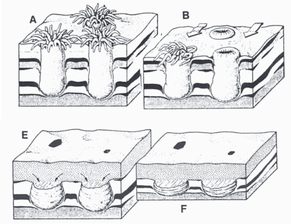
The Beltanelliformis appear at first glance to be similar to other disc-like Ediacaran forms associated with holdfasts. However, notice that the specimens pictures are in strong positive hyporelief, that is they extend quite some way out from the lower surface of the sandstone. This means that the sand infilled quite a sizable hole in the underlying mud. Notice also that, in the top two specimens, the fossil has a 'collar' around the basal disc, which makes the hole infilled by the sand cone-shaped, with the base being the smaller diameter part of the cone.

This configuration let to suggestions that the form represents the burrow of an anemone.

Below is a representation of that interpretation (from Schopf et al.).

However, more recent interpretations have Beltanelliformis as an algae.

What!? Algae!?

Yes, algae. Here's why.

The uppermost Doushantuo Formation (590-555 Ma) at Miaohe in the Yangtze Gorges area, is a series of dolomites that provide a rare Burgess-Shale-type taphonomic window on the Ediacaran. Within the Formation are found numerous carbonate compressions of organisms. This represents a different mode of preservation to that of the more typical sandstone preservation, at Miaho the forms are flattened carbonate compressions. Amongst the forms present are several that are interpreted as Beltanelliformis

Beltanelliformis brunsae from the Doushantuo Formation. Scale in 2, 1 cm. from Xiao et al. 2002)
The non-overlapping nature of the fossils is a good indication that we are dealing with benthic (bottom-dwelling) forms and not free-swimming forms that have died and sunk to the bottom. It may well be that the forms were actually tethered to bottom with a small holdfast structure.

The Doushantuo specimens have thing flexible walls. However, given the original specimen at the beginning of this post, it's clear that they would need to retain the three-dimensional shape during burial by sand.

A modern form that appears to fit the bill is the caulerpalean green alga Derbesia (photo credit).


Derbesia is benthic, is attached by a minute holdfast, and has the right shape and size. Also, it is organized as a coenocytic film of cytoplasm within a elastic and strong wall and surrounding one or more large, fluid-filled vacuoles. These vacuoles would be sealed and so couldn't empty on burial like holdfasts could. This makes it strong enough to make an impression in sandstone preservation!

So Beltanelliformis has gone from Cnidarian -> holdfast -> anemone burrow -> algae with holdfast.

Interestingly, apart from Nimbia and Aspidella mentioned in a previous post, Beltanelliformis has one of the longest time ranges, having been found in the pre-Ediacaran Cryogenian in Canada.

This again raised the possibility that several of the generic Ediacaran disc fossils could be something other than metazoans. That's actually fine. We have enough evidence that some of the discs represent metazoans. That some don't, should be expected. After all, if the Ediacaran assemblages represent vibrant, healthy ecosystems, they would be expected to contain microbial, algal, and metazoan, life.

Monday, January 3, 2011

To Epirelief or Hyporelief, That is the Question . . .

My last post on the possible 770 million year old Ediacaran fossils from Kazakhstan prompted some comments regarding epi- and hyporelief (go read the post and comments).

Basically, Ediacaran fossils are usually found on the underside of the sandstone bed in either positive relief (sticking out from the surface) or negative (sunk into the surface) hyporelief (hypo- is sciencey for 'on the underside'). This is dictated by the mode of preservation (again see the original post), and means that we do not find them on the top surface of the sandstone bed, or in epirelief. (Actually we do find them in epirelief, but only at Mistaken Point in Newfoundland where they have been mantled by volcanic ash - but that appears to be a one off and so does not represent normal preservation).

As an example here is a slab I collected from the Flinders Ranges in South Australia. First the upper (epi) surface.


As you can see, its rippled - strongly rippled actually - which puts pay to the ideas that the Ediacaran faunas were deep water, but that's another story. However, what the top surface isn't, is fossiliferous. There are no fossils on it.

Now lets take a look at what's underneath.


Ta Da! This was taken in situ, about 5 minutes after I'd found it. The photo is a bit blurry 'cos my hands were stili shaking!

Notice the smooth lower surface. This is because it is mantling a mud which does not form ripples. Also notice that there are three large discs in positive hyporelief and a Dickinsonia in negative hyporelief. What do you mean no!?. Ok, here's an annotated version.


The question has been asked why the counterpart of a positive hyporelief isn't a negative epirelief on the bed below. In other words if the fossil is formed by sand flowing into a depression in the underlying mud to form a positive hyporelief, should there also be a corresponding negative epirelief on the top of the underlying bed?

The answer is yes, but we almost never find them. The reason is that the underlying bed is made of mud and so compresses during diagenesis which disrupts the fossil. However, the main reason is that the mudstone weathers much more readily than the sandstone and forms a very friable, crumbly rock that simply weathers away.

We do have some examples of underlying epirelief fossils, but they are very rare. To find them you have to excavate the sandstone with the underlying mudstone still in place. Even then, they are very fragile. Normally this never happens in nature as the mudstone erodes away. The fossiliferous sandstone then weathers out and falls downslope to be found by palaeontologists.

Below is a typical Ediacaran location. The fossils are eroding off the top of the hill and can just be seen in outcrop. but the fossils are found on the scree slope, as float - that is, material that has eroded out of the outcrop and has slid downslope


So the answer is, yes there are there epirelief fossils but we almost never find them as they have weathered away. But they would be in mudstones and not in sandstones.

Sunday, January 2, 2011

770Ma Ediacara (?) Fossils from Kazakhstan (sadly no)

ResearchBlogging.orgThe Ediacaran Period represents that youngest part of the Proterozoic, and is famous for the first appearance of multicellular, metazoan fossils. The Period starts at the end of the great neoProterozoic Cryogenian, or glaciations - commonly called "Snowball Earth" - at around 635 million years ago, and ends at the start of the Cambrian (and Phanerozoic) at around 542 million yeras ago. While a few Ediacaran stragglers may appear in the Lower Cambrian, no unambiguous multicellular/metazoan fossils have been found below the base of the Ediacaran Period, despite several possible candidates (e.g here and here).

In a new paper, Meert et al. report the possible occurrence of Ediacaran fossils from 760-770 million year old Cryogenian rocks of Kazakhstan. If correct, this would extend the fossil record of metazoans back another 100 million years. Now, there is some evidence to suggest that metazoans were around in the Cryogenian, if only at the sponge-grade of organisation, but this is from biochemistry, not body fossils. So this would be the oldest metazoan body fossils yet found.

Two questions to be asked then. Are these deposits c. 770 million years old? Are these specimens examples of Ediacaran fossils?

I think the answer to the first question is yes, and the answer to the second question is no. I'll explain below.

Age
I have to say that Meert et al. have done an excellent job of mapping and correlating what appears to be a very difficult sequence. It's condensed and so quite thin (by contrast, the Ediacaran type section in Australia is several kilometres thick), a significant portion missing, and the area has been subsequently affected by tectonics.

Stratigraphic column for the Lesser Karatau sequence showing the relative locations of the fossil discoveries. The fossils under discussion are from the Upper Riphean Kurgan Formation (R3) (in brown) and the glacial deposits (in yellow) and cap carbonate represents the end of the Cryogenian glacial episode and the start of the Ediacaran Period. (Meert et al. 2010)
Now with a section like this it could be argued that the glacial rocks and cap carbonate represent glacial activity known to occur in the Ediacaran and that the underlying rocks are, in fact, Ediacaran. But I think Meert et al. have compiled enough evidence to support their stratigraphy. This includes radiometic dating of two separate samples from the Kurgan Formation that support a c. 770 million year age, as well as a δ13C stable isotopic curve that fits their interpretation (given the fragmentary nature of the curve, it could fit a number of scenarios, but it does fit this one.)

So notwithstanding the difficulties of the section, the date appears well supported. We appear to be dealing with c. 770 million year old rocks.

Fossils
1) Disc and stalk
Meert et al. have found structures in the c. 770 million year old Kergan Formation which they suggest may be referable to the Ediacaran forms Nimbia and Aspidella, as well as a second line of evidence, a possible stem extension emanating from the discoid fossil, and ask the question, are these Ediacaran fossils? As I said, I think the answer is no.

To show why, firstly I need to explain a bit about Ediacaran fossil preservation and positive/negative epi/hyporelief.

Ediacaran fossils occur primarily as marking on the surface of sandstones. These surface markings can be of two types, positive - standing up from the surface like a pimple, or negative - a depression in the surface like a dimple. Also the sandstone has two surfaces, a top surface - the epi-, and a lower surface - the hypo. Thus when we talk about surface structure, or "relief" - we talk of structure on the top bedding surface - epirelief, and structure on the lower bedding surface - hyporelief. So positive epirelief is a raised area on the top surface and negative hyporelief is a depression in the lower surface.

This is important because Ediacaran fossils occur as positive or negative relief on the bedding surfaces, but almost always as positive or negative hyporelief - that is, on the bottom surface.

The reason for this is that preservation is by sand covering a surface that has animals on it. The sand (which will eventually turn into sandstone) covers the animals. Where the animal is tough, it will push into the overlying sand, causing a negative hyporelief impression (of the top surface of the animal) into the bottom surface of the sand. Where the animal is buried in the underlying sediment, such as holdfasts, the sand flows into the depression caused by the collapse of the holdfast during burial, causing a positive hyporelief impression.


The example above is of the lower surface, The Form D projects out from the surface and so is a positive hyporelief impression (the overlying sand has pushed down into the Form D stolon structure to form the impression. By contrast, the Parvancorina has pushed up into the overlying sand and so is preserved as a negative hyporelief impression.

OK, lets deal with the second line of evidence first - the possible stem extension emanating from a possible discoid fossil.

(A) Discoidal impression (negative relief) with possible stem-like extension. (B) Sketch showing the location of discoidal fossil and possible stem. Meert et al. 2010.
Just to be clear about what is being discussed, here (at left) is a modern sea pen (photo credit) we can use as an example of the frondose forms around in the Ediacaran. The structure half-buried in the sediment is the disc-shaped holdfast. Underneath the holdfast is a thin tube - the peduncle - which acts like the foot of a clam and aids in digging the organism into the sediment. The stem and frond extend out into the water column and the whole thing is held up by water pressure. The organism can inflate by pumping water into the holdfast to allow it to act as an anchor, and also into the stem and frond to hold them rigid. Pumping water out deflates the organism. Modern sea pens will deflate on touching, and collapse down onto the sediment, as a method to avoid predation or damage.

According to Meert et al., their find may represent a holdfast and part of the attached stem that has made an impression in the sediment, in negative relief. The authors do not say if the surface is the top or bottom (and they may not know if the sample was collected loose and not found in situ.

However, holdfast impressions are almost always found as positive hyporelief impressions, i.e. they stick out from the lower surface. They are not found as negative relief, i.e. as depressions, as the Kurgan Formation specimen is. To find out why that is, let's look at an example.

Three-dimensional positive hyporelief fossil of a holdfast and stem from the Ediacaran of South Australia. HF impression of holdfast, IS intermediary surface within the sandstone, LS lower surface of sandstone, P peduncle, S impression of stem, W wrinkles in top surface of holdfast. White arrow is the direction of current depositing the sand.
Above is an actual Ediacaran holdfast and stem fossil. There's a lot going on here (compared with the Meert et al. specimen where no structure is present) so it'll take a bit of explaining.

Firstly observe that this is in positive hyporelief - it's on the bottom surface of the bed. As I said, pretty much all holdfast fossils with stems are positive hyporelief. This is because the organism is sitting in the underlying sediment and gets inundated and covered by a mantling sand deposit which will become the overlying sandstone bed. Since frondose forms will deflate on contact with the enveloping sandstone, the organism pumps out water and collapses. This means that the stem falls over, usually in the direction of sand transport. The stem often gets trapped within the sand and rarely comes to rest on the sea floor because the sand has started to deposit as the organism responds. This normally results in the stem coming to rest at an shallow angle to the sea floor/base of the sand body - that is, it sticks up at an angle into the sand rather than laying down flat. This can be seen in the Ediacaran example shown above. The stem impression (S) can be seen heading off at the ten o'clock direction but it is obviously heading off into the sandstone - the edge of the stem impression is lower in the sandstone than the impression of the holdfast, and, as this specimen is shown upside down, the stem in heading upwards into the overlying sandstone.

The wrinkling texture is the upper surface of the holdfast reacting to the stem being pushed over and collapsing and shows that the holdfast stayed in place during burial.

The organism is now covered by a layer of sand, with the stem trapped within the sand at a shallow angle. As the water has been pumped out, the holdfast will no longer stay inflated and so the holdfast upper surface sinks down onto the lower surface causing a shallow depression. The upper surface also takes on the shape of the lower surface where the hollow peduncle is, creating a ring shape (P). The overlying sand then flows into the shallow depression and takes on the shape of the combined lower and upper holdfast surface structures in a process called gravity casting. Also, as the stem is angled upwards, the sand with collect behind it and be trapped between the stem and the holdfast. We can see this is the example above. The sandstone covering the stem has been removed, leaving a lighter coloured sandstone. The stem starts out on top of the holdfast, but ends up on a lower, intermediate surface (IS) (lower from our prespective, higher when the specimen is rotated to the correct orientation).

What you end up with is a positive hyporelief impression of the combined upper and lower holdfast surface, plus the base of the stem. Furthermore, this is the only way this can be preserved. We tend not to see holdfast impressions on the tops of sandstones because the organisms appeared to favour softer, finer sediments such as silts and clays, and where active microbial mats occur.

With that in mind lets look at the Meert et al. specimen again.


This time I've annotated it to mark out the various levels in the specimen, with A the highest, B an intermediary level, and C the lowest (assuming we are looking at the top surface, or the reverse if we are looking at the lower surface).

First of all, this is a negative relief. As I explained above, holdfast and stem preservation is usually by gravity casting which produces positive relief. If we are looking at a bottom surface, maybe the organism remained intact and 'pumped up', forming a hollow in the overlying sand. But in that case the stem would also remain intact and we wouldn't see the impression of the stem in the resultant fossil. So if the stem is represented, then the organism had collapsed, in which case the holdfast would have collapsed and we would get a positive hyporelief as notmal.

The structure can't be a positive hyporelief that has eroded out the produce the depression because the original fossil is just a surface impression. The sandstone underneath the fossil is the same sandstone the rest of the rock and so there can be no preferential weathering to produce the hollow.

If we are looking at a top surface, then it is unlikely to be a holdfast and stem as we do not find them on top surfaces of sandstones. Even if it were, the stem appears to have been pushed into the sand body to a level equal to, or even greater than, the depth of the holdfast. Again an unlikely occurrence, as a slight impression on the underlying sand is the most you are likely to get.

Given that this is a negative relief and the lack of any distinguishing structure, it is most likely that the specimen is simply an erosional feature and not a fossil.

2)Nimbia and Aspidella
Specimens referable to Nimbia and Aspidella are reported from the c. 770 millio year old Kurgan Formation.
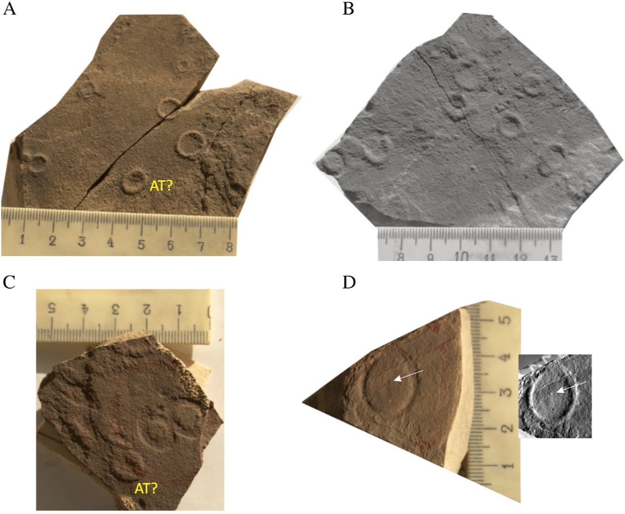
Our specimens were discovered in a brown-red shale within the largely siliclastic Kurgan Formation and are preserved in both hypo and epi-relief. They are circular to oval-shaped impressions that surround either a smooth interior or circular to oval indentations with a smooth interior. (Meert et al. 2010.)
(A) closeup of the Nimbia fossils in positive epirelief, AT marks the occurrence of a possible invaginate morph of Aspidella terranovica; (B) B&W photo of Nimbia impressions shown mostly in positive epirelief; (C) Nimbia fossils in negative relief with a possible invaginate morph of Aspidella terranovica in positive relief; (D) large c. 2 cm (long axis) c. 1.5 cm (short axis) negative hyporelief impression of Nimbia occlusa and an ‘inversion’ of the photo the fossil might appear in positive epirelief. White arrow points to a possible raised central nodule observed in Nimbia occlusa fossils elsewhere. Meert et al. 2010.
Taking Aspidella first. Actually, attributing the form to Aspidella doesn't tell us much. This is because Aspidella should now be considered a form genus - that is a grouping of similar forms (i.e. discs) and not representative of a single organism, or related group of organisms. In other words, a lot of different things make disc impressions, e.g. algae (algal biscuits), microbial colonies, fungae and metazoans. Calling the specimen Aspidella simply says that it is a certain kind of disc, not that it is the Ediacaran form Aspidella terranovica. There are a lot of morphologies within the Aspidella group and they are not produced by the same organism, or even closely related organisms. This means that more than one organism is included under Aspidella. Even worse, no consistent distinction can be made between the various disk structures, making it difficult to accurately assign a particular disc to a particular organism. Where other evidence exists, i.e. stem impressions, ornamentation, etc, it is possible to identify individual organisms or groups of organisms. But simple discs remain probematic. Being a disc does not make it a holdfast!

It is entirely likely that Aspidella actually represents a group of diverse, not closely related, organisms - microbial, algal, fungal and metazoan - that produce discs and overlap each other to give Aspidella a large time range, but this time range is not for a single species or even genus.

This boils down to the fact that Aspidella with stems or other ornamentation, and associated with other Ediacaran forms, can be considered a metazoan fossil. Other Aspidella with no other ornamentation, and not associated with Ediacaran forms, could be anything.

The Nimbia find is in a similar situation. Do we have Nimbia? Yes. But here also, I think that the form Nimbia, like Aspidella, may hide a multitude of sins, or at the very least a number of different organisms. Except I think we have proof here that Nimbia is not an Ediacaran metazoan - or at least this Nimbia is not an Ediacaran metazoan.

The specimens of Nimbia from the Kurgan Formation are found as positive epirelief, negative epirelief, and negative hyporelief. This does not happen for Ediacaran fossils, as discussed above. Since these Nimbia show a number of modes of preservation, and they all show the same structure regardless of mode of preservation, it is apparent that the form that produced it is rigid. It has to be if it produced the same shape whether on the top surface creating a positive or negative imprints, or on the bottom surface. The fossil is the same independent of preservational style, and so whatever is causing the fossil must therefore be rigid like a coin. That rigidity, and the various preservational styles, means that it cannot be a holdfast.

It has been suggested that Nimbia represents microbial colonies. Hard, well agglutinated disk-shaped colonies would be tough enough to cause the impressions, and some have been known to develop a central nodule similar to that found in some Nimbia. This would explain the similar shape through different modes of preservation, and the large time range of the form.

Finally, Ediacaran fossils are normally found as assemblages, with a number of different forms present. The Kurgan Formation specimens are of one main type, Nimbia, and a few Aspidella. Too restricted to be considered an assemblage (a random collection would be expected to uncover more than two forms if an assemblage was present). Even if this find represented an early stage in the evolution of metazoa, it would be expected that more than two forms would be present.

So, discoial fossil with stem? - no. Aspidella? - yes, possibly, but that doesn't mean it's an Ediacaran or even metazoan. Nimbia? - yes, but probably not metazoan.

In properly posing questions about their finds instead of making pronouncements, and suggesting a number of possible interpretations for their finds, Meert at al. have highlighted the important implication of their finds.
It is possible that our discovery of Nimbia occlusa and Aspidella terranovica(?) in sedimentary rocks during the early Cryogenian (N766 Ma) lends support the alternative hypotheses regarding these fossils and remove them from consideration as true metazoa(Meert et al. 2010.)
I think they are right, they have provided evidence that the forms are not metazoan - or at least not "Ediacaran" (as in found amongst Ediacaran assemblages) - fossils, and that the simple disc-shaped forms found in the Proterozoic comprise a form group with contributions from a number of metazoan and non-metazoan sources.


Meert, J., Gibsher, A., Levashova, N., Grice, W., Kamenov, G., & Ryabinin, A. (2010). Glaciation and ~770Ma Ediacara (?) Fossils from the Lesser Karatau Microcontinent, Kazakhstan Gondwana Research DOI: 10.1016/j.gr.2010.11.008


Further reading on Ediacaran discs

Gehling, J.G., Narbonne, G.M., and Anderson, M.M (2000) The first named ediacaran body fossil Aspidella terranovica. Palaeontology, 43: 427-456. DOI: 10.1111/j.0031-0239.2000.00134.x

MacGabhann, B.A. (2007) Discoidal fossils of the Ediacaran biota: a review of current understanding. Geological Society, London, Special Publication, 286: 297-313. DOI: 10.1144/SP286.21

Sunday, September 19, 2010

Proterozoic Sponges Claim Doesn't Hold Water

ResearchBlogging.org


The geologic account of ancient life is plagued with reports that do not withstand critical assessment. This is a special problem in the older rocks, where reports of spurious records continue to dilute the authentic record of evolution on the primitive Earth . . .

. . . It is left to the reader to draw conclusions about similar instances not here alluded to; the record of alleged pre-Phanerozoic life is full of them.
Preston Cloud (1973)

Harsh perhaps, but Preston Cloud's words should be engraved on the cover of every Proterozoic geologist's field notebook.

Frankly, the Proterozoic is weird. Most of what you think is organic, isn't. The vast majority of the rest is microbial mats. And the stuff you really, really think could be metazoan, is usually microbial mats playing silly buggers.

I don't want to become a party pooper (and no that's not a plug for Bora's pootopia). I don't, I really don't.

I want to be able to yell and scream about some new find that pushes our knowledge of the early evolution of metazoans back well into the Proterozoic. I do, I really do.

But I can't.

In Nature Geoscience, Maloof et. al discuss possible sponge-grade metazoans from the approx. 640 million year old Trezona Formation in the Flinders Ranges of South Australia.

The Trezona Formation just underlies the Marinoan glacial deposits (dated to approx. 635 million years ago) which mark the final throw of the of the Cryogenian dice, and are overlain by rocks of the Ediacaran Period. The Trezona marks the onset of the Marinoan glaciation and contains shallow water stromatolite flake breccia and bioclast packstones filling gaps between stromatolite heads:
. . . we identified a great diversity of bioclasts. Most packstones contain clasts of probable microbial origin, such as spalled flakes of adjacent stromatolite laminae and ripped-up and rolled-up sediments with cohesion enhanced by the presence of microbial mats. However, many bioclasts have anvil, wishbone, ring, and perforated slab morphologies that are difficult to assign to an abiotic roll-up or bacterial mat origin. in addition, the red colour and calcite composition of these distinctively shaped clasts are unique to the packstones (and even packstone clasts entrained in the overlying Elatina Fm diamictite as far as 65km from the nearest Trezona Fm stromatolite reef outcrop) and are not found in situ in the layers elsewhere in the Trezona Fm that could have been brecciated and transported. Therefore we suspect that the 1-cm-scale red bioclasts represent the remnants of a community of organisms endemic to the stromatolite-packstone environment. (Maloof et.al 2010.)
This is what we are talking about

Outcrop photos from the Trezona formation showing the range of clasts
including "anvils" (d) and "perforated slabs" (g). (Maloof et.al 2010.)

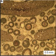
2D thin sections show the clasts are composed of a mixture of opaque clays, quartz grains and micro-crystalline calcite, finer than the surrounding matrix, with a sharp, continuous contact with micritic rims

Thin-section photomicrographs from the Trezona formation. (Maloof et.al 2010.)

The authors then describe 3D shapes using serial sectioning (grid a bit off, photograph, grind a bit off, photograph). These 3D images show a three-dimensional network of 1mm diameter interconnected tubes. The tubes are also lined with micrite.

Three-dimensional reconstruction of the Trezona Formation
structures. (Maloof et.al 2010.)


So we have stromatolites with infilling packstones contain a diverse set of bioclasts representing chips from associated stromatolites and ripped up and rolled sediments that have been glued together by microbial mats. However, the paper claims that some of these bioclasts have shapes, include anvil, wishbone, ring and perforated slabs, that cannot be caused by nonbiological actions for the following reasons:

1) The shapes cannot be formed by rock chips
2) The red colour and micrite composition is unique to the deposits - no source of the chips can be found.
3) The micrite coating in the tubes and around the clasts may represent weakly calcified cell layers.
4) The interconnected 3D tube structures and the 3D symmetry support a sponge hypothesis

1 and 2
OK, lets split these up. The first two deal with the lithology and composition of the clasts. The authors say that the clast shapes produced cannot be formed from mud chips or bits of stromatolite, and that is true. It is also true that there are no lithologies similar in colour or composition to the clasts anywhere else in the Trezona Formation. This means that the shapes cannot represent chips eroded off earlier formed mudstones because there are nor similar lithologies or rock types in the area, and even if there were, rock chips cannot create the shapes seen. But there are problems with using that as supporting evidence for a biological origin. There are other explanations. Other abiogenic explanations.

The local explanation for the clasts is that they are mud flakes not mud chips, that is, not mud chips eroded off pre-existing rocks, but mud flakes formed when muds are deposited in ephemeral or short-lived deposits, such as ponds after flooding surrounding areas, or overbank deposits.

Drying mud Gammon Ranges. Photo Credit.

These muds settle and dry out and can form all sorts of shapes.

Drying mud, Canyonlands National Park. Photo by Douglas.

In some instances the mud flakes can completely roll up

Drying mud from Death Valley. Photo by Don Gale.

Check out one of the rolled mud flakes pictured above compared with a couple of the clast shapes


Clearly some mud flakes can take on the shapes that mud chips cannot, and can form some of the shapes present in the Trezona Formation that are being used as evidence for a biologic origin.

But it gets worse. See the arrowed clasts in the image with the "anvil" shape (at right). These are clearly mud flakes. They show a common curled profile that can be seen in the examples of recent drying muds above. Nor do they contain the tubes that are supposed to be a feature of the "sponge" clasts.

Now, here's the interesting bit. Both the obvious mud flakes and the "sponge" clasts are the same colour AND have the same composition (opaque clays and microcrystalline calcite, with the occasional very fine quarts grain - see photo-micrographs above) in other words your typical mud overbank deposit. Especially since the Trezona is pretty much a lowstand deposit (lower sea level). Some of the limestones deposited prior to the Trezona Formation, when sea levels were higher, are now exposed, providing a source for carbonate and putting some distance between the Trezona depositional environment and any non-carbonate source material (meaning that only very fine grained material is likely to reach the depocentre).

I think it is clear that the red clasts are mud flakes from overbank deposits, rolled up and redeposited between the stromatolites. In other words, waters carrying red carbonate mud, overtop riverbanks and settle into ponds. The mud settle out and the ponds dry up. The mud then dries, with the carbonate providing a good cement. The mud cracks, curls and eventually are dispersed by winds rain or floods into the nearby sea. The mud could be sourced from further inland on the Gawler Craton, and the carbonate sourced from limestones deposited during the period before the current regressive cycle, of which the Trezona Formation represents a lowstand or low sea level phase. The ephemoral nature of these overbank deposits, and their lack of lateral extent means that they do not contribute to the rock record, but dry out, crack, curl and are dispersed by wind and/or water. This also explains the occasional presence of "silica blebs" within the clasts. These represent very fine sand grains incorporated into the muds as they are deposited.

Clearly the mud flakes and "sponge" clasts have the same source - which is not organic. The shapes and colour cannot be used as supporting evidence of an organic origin for the clasts.

The authors suggest that the red matrix could be a replacement:
Alternatively, the original organic skeletons could have been coated in a bacterial extracellular polymeric substance following their death. The chemical composition of the Fe, Na, K clays found in the Trezona Fm bioclasts is similar to that of augenic minerals precipitated by microbial biofilms during the replacement of soft tissues. The extracellular polymeric substance would have also formed a template for abiotic calcium precipitation. (Maloof et.al 2010.)
Now if I'm reading that right, it looks like the authors are suggesting that the original skeleton of the "sponges" has been replaced by the red mud, since there is no evidence for spicules or any other type of a primary skeleton.

There are two problems with this explanation for the absence of a primary skeleton. The first is the fact that the red matrix composition is shared with clasts that are clearly mud flakes. The second problem goes to number three in their list of supporting evidence, that the micrite coating in the tubes and around the clasts may represent weakly calcified cell layers.

3
The authors comment that the "sponge" clasts are surrounded by a micrite rim (the dark outer rim around the clasts in the photo-micrograph at right. I don't know why they don't use the standard terminology - micrite envelope), which is also present lining the tubes. They suggest that:
the micrite of uniform thickness and texture coating both the exterior surface and interior canal walls could represent weakly clacified cell layers sandwiching the mesohyl of a sponge grade organism. (Maloof et.al 2010.)
To their credit the authors then demolish this argument by pointing out that:
However, texturally similar (but less uniform thickness) micrite also coats peloids that do not seam to be part of the Trezona Fm organisms.
Yes, that's a bit of a problem. It's a bit difficult to suggest that the micrite envelopes are evidence of mesohyl when almost everything in the deposit has them.

Micrite envelopes are well known and are generally considered to be formed from filamentous organisms (bacterial, algal or fungal) who's filaments calicify and coalesce into a intertwined mesh around the clasts. In this case the areas between stromatolites are colonised by numerous filamentous organisms that grow around all the clasts present and eventually form the micritic envelopes. So having micritic envelopes around the "sponge" clasts and within the tubes cannot be used as evidence of sponge-grade tissues.

But there's another problem.

As stated above, the authors suggest that the mud matrix could be a replacement for the original skeleton structure around the tubes. The presence of the micrite envelopes is really problematic here. The authors comment that:
However, the contact between micrite coating and mixed clay-chert-calcite interiors is usually sharp, with no evidence of diffusive or porosity-following micritization. (Maloof et.al 2010.)
This means that any replacement of the original skeleton must have occurred prior to the micrite envelope being laid down. However, the tubes must have been sealed during replacement as there is no replacement material in them. After replacement, the tubes must have opened again to allow the micrite envelope to form.

Unlikely.

The evidence suggests that the red matrix is not a replacement, but a primary feature.

4
Which brings us to number 4, that the interconnected 3D tube structures and the 3D symmetry support a sponge hypothesis. I'll say up front that I do not know what the tubes are. They could be sponge tubes, but the evidence for that is equivocal at best.

A more likely explanation is that the "sponge" clasts are mud flakes that have been coated by filamentous organisms, rolled around and glued together. The tubes would then be where the filamentous outer coating of individual clasts, acting as a buffer and holding the clasts apart as a number of clasts were bound together.

It is interesting to note that an analysis of similar lithologies from rocks of the the equivalent Cryogenian interval south of Adelaide found;
. . . a light greyish microcrystalline limestone in which numerous flakes of grey calcareous mud are set. The flakes are typically intraformational and a gradation is noticed from intraformational breccia to edgewise conglomerate. Most of the flakes are flat, but there is a tendency to turn at the edges; length varies from several to 20 mm. in thin section or on polished faces there is a superficial resemblance to annelids (reference), but this is quite lost when the third dimension is considered. (Sprigg 1942)
In the Proterozoic, weirdness rules. This makes interpretation difficult. I don't think the evidence presented in the paper is enough for a sponge interpretation to hold water. However, we've been misinterpreting the Proterozoic for some time, and the authors are in good company.


Cloud, P. (1973) Pseudofossils: A Plea for Caution. Geology, v. 1, p. 123-127.

Maloof, A., Rose, C., Beach, R., Samuels, B., Calmet, C., Erwin, D., Poirier, G., Yao, N., & Simons, F. (2010). Possible animal-body fossils in pre-Marinoan limestones from South Australia Nature Geoscience, 3 (9), 653-659 DOI: 10.1038/NGEO934

Sprigg, R.C. (1942) The Geology of the Eden-Moana Fault Block. Transactions of the Royal Society of South Australia, 66(2), 184-214. Download

Saturday, August 7, 2010

2.1 Ga Multicellular Colonial Organisms - Update 2


In posts here and here I discussed why I thought the 2.1 Ga structures from Gabon, figured in the Nature paper, were actually microbial mats and not examples of multicellular colonial organisms.

I now have some further info which strengthens my view. I'd like to thank Dr Diana Cuadrado of the Instituto Argentino de Oceanografía who very kindly sent me some more images of microbial mats.

This mainly revolves around the claim in the paper that microbial mats . .
. . often leave characteristic in carbonate ad siliciclatic rocks. Such structures, however, including those formed in shales and mudstones, do not resemble the Gabon fossils". (p. 103)
As I've discussed previously, I disagree.

At the top of this post is an example of a modern microbial mat (top image), compared with a photo of the 2.1 Ga structures (bottom image). The black box in the top image represents the area of the lower image at the same scale (The bar scale is 10 cms in the top image and 1 cm in the bottom image). This reinforces just how small these 2.1 Ga structures are.

Here then, is more evidence that microbial mats can produce the structures seen in the 2.1 Ga specimens.


Here is a close up of a large gas bubble in a modern microbial mat (it was taken last week - you can't get much more modern than that!). As you can see by the comparison with one of the 2.1 Ga specimens at the same scale, gas bubbles in microbial mats are on the same scale as the 2.1 Ga specimens.

For me this is the clincher (if a clincher were needed)


The top photo is of a modern, ruptured gas bubble, the bottom is of a 2.1 Ga specimen. See how the modern microbial mat is flexible enough to fold and stay intact even when torn. But, see the folding to the top and right of the hole. It's almost an exact match for one of the 2.1 Ga specimens figured, to scale, below. No that's not quite right . . . it's an exact match!

Not only that but there is a small fringe beyond where the folds end in the modern example, similar to that seen in the 2.1 Ga specimen.

To say something like "Ta Da" at this point would be churlish, juvenile and unprofessional, . . . so

TA DA!

But wait, there's more.


This is a photo of a mature microbial mate with a couple of overturned pieces. Not the cracking in the larger piece around the margin. his may be the cause of the "radial fabric" of the 2.1 GA strucutures. The smaller piece has even more marked cracking and looks similar to this 2.1 Ga specimen below.


This latest evidence strengthens the argument that the 2.1 Ga structures are pyritised microbial mats and not multicellular colonial organisms.

Tuesday, July 13, 2010

2.1 Ga Multicellular Colonial Organisms - Umm, Not (update)

I previously discussed why I don't think the 2.1 Ga structures found in Gabon are multicellular colonial fossils.

Having looked at the Nature paper some more, another thing springs to mind.

Scale.

Here are some of the structures as figured in the paper.


Note the scale bars against each example. Those scale bars represent 5 mm! That's right millimetres!

These things are small. Especially the central area that contains the folding.

Now, compare that with the examples of bubbles in microbial mats

Photo from Cuadrado and Pazini (2007)

There is no scale, but some of the bubbles must be at least the size of the central folded area of the specimens figured in the Nature paper. This means we have structures documented today in microbial mats that equate in size to the central area of the structures from Gabon.

Now, what would happen to a gas or fluid filled bubble if it were covered with sediment and the gas/fluid escaped? It would deflate. Not deflate flat, as the microbial structure would be too thick. Maybe it would fold on itself just like the central structure in the specimen in row two above? Especially if there was an encouraging push from overlying sediment.

I'm even more convinced that the Gabon structures are microbial mats.

UPDATE 2

Diana G. Cuadrado and Natalia V. Pizani. (2007) Identification of microbially induced sedimentary structures over a tidal flat. Latin American Journal of Sedimentology and Basin Analysis. v.14 n.2 La Plata ago./dic. 2007

Monday, July 5, 2010

2.1 Ga Multicellular Colonial Organisms - Umm, Not


ResearchBlogging.org

2.1 GA structures on black shale bedding surface. Scale bar 1 cm. (El Albani et al. 2010)


The latest edition of Nature has a paper claiming 2.1 billion year (Ga) old multicellular colonial organisms from Gabon. This is a remarkable claim, since the oldest definitive large multicellular organisms so far found in the fossil record are from less than 600 million years ago (It is still not certain if the 2 Ga Grypania spiralis is a eukaryote or large bacterial colony). So remarkable is the claim in fact, that it made the front cover of Nature.

I think they are wrong.

Lets be more specific here. There are two claims - that the structures are 2.1 Ga old, and that they represent large multicellular colonial organisms.

First the date. That appears well constrained. Chris Rowan has good coverage of the dating at Highly Allochthonous, so I have no argument against the age.

I don’t think they are multicellular colonial organisms. I think they are pyritised remains of microbial mats, or structures associated with microbial mats.

There’s been ample coverage of the paper, but I want to concentrate on the reasons why the authors think that the structures represent organised colonial organisms.

The structures are found in unmetamorphosed black shales. Over 250 specimens have been recovered. They are pyritised and embedded within the sediment. They range from 7-120mm in length, 5-70 mm in width, and 1-10 mm thick. They can occur in densities of up to 40 specimens per square metre, at random orientations, but all are horizontal to bedding. All are wrinkled to some degree, with some having large central pyrite nodules. Some show significant folding. All show radial cracks. The authors used micro-computed tomography (Micro-CT) to produce three-dimensional images of the structures. They also undertook carbon and sulphur isotope analyses of the host rock and the structures.

On the following lines of evidence the authors decided that the structures represented multicellular colonial organisms:

  1. The structures shown under Mirco-CT are not the same as produced by microbial mats.

  2. They are unaware of any inorganic process that could produce the folding patters seen in the structures.

  3. The folding suggests an originally cohesive flexible sheet.

  4. They are unaware of any inorganic process that could produce the radial fabric seen in the structures.

  5. The radial fabric represents peripheral accretion of flexible organic matter.

  6. The 12C/13C carbon isotope ratio in the host sediment is different to that in the structures suggesting the structures represent distinct organisms.

  7. Steranes have been found in the shales, a compound associated with eukaryotes.

  8. A lack of support for any other inorganic origin.
I think pyritisation of microbial mats is a better explanation. Here’s why.

I’d like to take 1, 2, and 3, together because they all relate to the physical properties of microbial mats.

First let's take a look at what we're talking about.

Micro-CT-based reconstructions and virtual sections of four specimens from the FB2 member of the Francevillian Group. First Column - original. Second column - volume rendering in semi-transparency. Column 3 - Transverse two-dimensional specimen. Column 4 - longitudinal section running close to estimated centre of specimen. Scale 5 mm. (El Albani et al. 2010)

The structures are pretty much all pyrite. The absence of pyrite is marked by the radial cracks. The bright central area in rows 3 and 4 represent a central pyrite nodule. This is not present in all specimens. This is important, as the large thickness values for specimens are all associated with this central nodule. Away from the nodule, or when a nodule is not present (e.g. row 2 above), the structure is a thin film of pyrite around 1-2 mm thick.

This is really important. All the folding is just that, folding. The apparent holes (black areas) in the images above do not represent complex structure within the specimen. The image is a slice through the specimen at a certain level. Where the pyrite film is folded below the level being imaged (downward folds), or completely above it (upward fold), you get a black space. The images appear to show complex structure, but it is simply a line drawn across an undulating surface. There is no internal structure here other than the radial fabric.

Further, and critically, the authors have concluded that the thin pyrite film and the central nodule represent different periods of pyrite formation. This is supported by sulphur isotope data, and I am happy to accept their conclusion.

This means that the large nodules have little to do with the original structure of the specimens, and, in fact they appear to occupy a central cavity in the specimen when they are present. An example of an actual nodule is below.

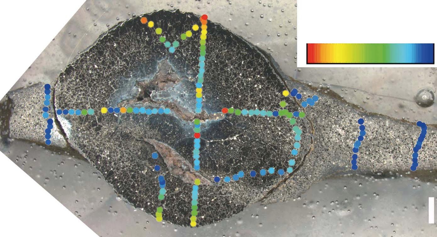
Section through a specimen showing central pyrite nodule surrounded on both sides by sheet material. Scale bar 1 mm. (El Albani et al. 2010)

In this specimen you can see that the nodule and the thin film on either side of it is almost all pyrite. The gaps in the center of the nodule are unexplained, but probably host sediment. Ignore the pretty colours. that refers to sulphur isotope analysis which I'm not questioning.

So, if we ignore the central nodules for the present, we are left with an original structure that is a thin film approx. 1-2 mm thick which has been thrown into folds in some specimens (e.g. row 2 above) but not in others (e.g. row 1 above).

The authors claim that the folding and the radial cracks cannot be accounted for by inorganic processes. I think they can.

Look! over there on the right! Is a 2.1 Ga colonial organism with a radial fabric? Is it a Proterozoic jellyfish? No, it's . . well, I'll explain later. But compare this example with rows 2, 3 and especially 4 of the Micro-CT images above. Notice that it has a central zone which is distinct from the periphery. Notice there is a faint dark band separating the central core from the periphery. Notice also the distinctive radial 'cracks' that spread out from the core Central mass? Check, radial cracks? Check. This would appear to be a good match to the 2.1 Ga structures.

But if you are not convinced, here's another variety of the same structure. this time we'll compare it directly with row 2 above as this has the best fold structure presented.


Can you see the fold pattern at the centre of the lower photos? It's quite similar to the fold pattern in the Micro-CT image. These images are of water escape structures in Ediacaran sandstones. The difference in colour you can see is due to the sandstone being deposited wet and dirty - i.e. waterlogged and with a significant amount of silt and clay (the red stuff). Escaping water carries the red silts and clays away with it, leaving the white sand behind.

Three things to note. They can produce radial structure, they can produce fold-like structures, and they are pretty similar in size even though they are from different sandstones.

Now, for the record, I don't think that the radial structures in the 2.1 Ga structures are water escape features (I've another idea for them), I'm just showing that such structures can be formed inorganically. Although I am suggesting that water escape could be an explanation for the folding we see in the row 2 specimen.

The authors discount water escape as an explanation because the underlying sediment shows no disruption such as that shown in my Ediacaran example viewed from the side. Plus, shales are not known for their permeability (ability to allow water to flow through it). But we are dealing here with microbial mats. Mats can act as a barrier to water or gas flowing below them. In such instances, rapid loading by waning storm sediments could cause water or gas trapped under the microbial mats to tear through the mat, creating some neat folding patterns and not disturb the underlying sediment too much.

Actually I'd better show you some mats so that you know what I'm talking about. Here's one.

A juicy, yummy microbial mat, full of cyanobacterial goodness from Yellowstone Park. Photo Carnegie Institution

(And yes I do consider Yellowstone to be a good analogue for the 2.1 Ga Proterozoic, because while the hydrothermal pools are aggressive to microbial mat grazers, so was the 2.1 Ga Proterozoic, because, well, there weren't any.)

Do you think the microbial mat above looks like what the authors have imaged? I do.

And just so we're clear on how flexible microbial mats can be, here's another photo.

Photo from Cuadrado and Pazini (2007)

Here are some more Microbial mats.

Photo from Cuadrado and Pazini (2007)

Photo A shows microbial mats draped (folded) over cracks. Photo B shows gas bubbles trapped under a microbial mat. What? you thought I just made that gas stuff up?!

But this is the clincher for me.

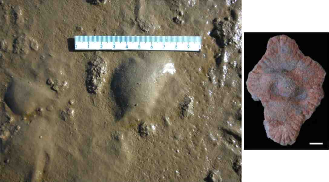
Microbial mats at West Chaplin Lake (Bowman and Sachs 2008)

There are a number of things to notice here. Firstly the mats are not one continuous sheet, but here are two discrete mats about the same dimensions as the 2.1 Ga structures. Also note the lower mat has been torn to produce a clean, sharp, high angled edge. This shows that these things can be torn without losing structure, so simple folding should be no problem. Speaking of folding, check out the left margin of the upper mat. See the fold? Remind you of anything? This maybe?


So microbial mats can fold and wrinkle without tearing. I agree with the authors that the structures represent originally cohesive flexible sheets, but microbial mats not colonial multicellular organisms.

There are other methods to fold or wrinkle microbial mats. Storms could rip up mats or partially dislodge them for example, and the authors interpret the environment as deltaic, influenced by storms. Elephant skin textures on bedding plains have been interpreted as being caused by wrinkling and folding microbial mats.

I hope this has shown that the structures seen in the 2.1 Ga specimens, the folding and wrinkling, can be produced by inorganic means on biological sheets without invoking a new class of organism.

OK, on to numbers 4 and 5, the radial fabric.

This is one of the main arguments for the structures being a colonial multicellular colonial organism and it certainly is striking. but, as I showed above, radial fabrics can be caused by other things. inorganic things.

This irregular radial fabric, along with the the fact that it is commonly deflected to meet the edge of the structures is taken as evidence that the structures grew by peripheral accretion of flexible organic matter.

However, there are a few specimens where this radial fabric appears to go right through the structure.
Scale bar 1 cm

In one example, the fabric appear to actually cut through the central nodule

Scale bars 5 mm


How could a growth-related fabric cut through a nodule that wasn't there until late in the diagenetic process?

I think it's down to water again.

Lets assume that the mats/colonial organisms have gone through initial diagenesis and have been converted to pyrite. As diagenesis proceeds and load on the sediment column increase, the mudstones start to compact. Muds can contain 60% water which means that they have a long way to compact. Problem. Pyrite sheets can't compact.

I think that as compaction progressed and the pressure increased, the thin outer margin split in a regular pattern. As the process continued, the cracks spread further into the body of the specimens and sediment would be forced into the cracks. As we have an example of crack through the central nodule, it may be that this cracking occurred late in the diagenetic process, when compaction was reaching it greatest extent.

So rather than be evidence of peripheral growth, the fabric may be diagenetic.

Number 6 and 7 are linked, 6 is the carbon isotope ratio, and 7 the presence of steranes.

The authors found a difference in the organic carbon isotopic composition between the host rock and the structures. This, they say, shows that the structures were distinct organisms.

Without going into too much detail, carbon is present in two main isotopes 12C and 13C with one extra neutron in the nucleus. Organisms preferentially take up 12C in different amounts depending on their metabolic pathways, causing a shift in the 12C/13C ratio. Measuring this difference or delta 13C can help identify the origin of the organic mater.

The host rock has a delta 13C value of -27 while the structures had a value of -32. Now -27 is roughly an average value for eukaryotes, and so would be expected to represent the fallout from the water column into the sediment of dead eukaryote plankton. A value of -32 is more like bacterial signature. This would tend to support my suggestion that these structures are pyritised microbial mat fragments.

Number 7 is the observation that steranes have been found in the shales (note the shales not the structures). Steranes are compounds associated with eukaryotes. But if the explanation for the delta 13C values is correct, the steranes would also be produced from the eukaryote plankton and can't be used to support the suggestion that the structures are eukaryotes.

Finally number 8, a lack of support for any other inorganic origin.

I think I've put up enough suggestions for inorganic input into the formation of these structures. but here is a rough explanation:

- Microbial mats grow on delta front muds - probably as individual round patches, possibly connected by thin connections to other mats.

- Storm activity rips up or distorts the mats into folds and waning storm currents deposit a layer of sediment over them.

- Pore fluids equilibriate and the mats are surrounded by sulphide dominated pore fluids.

- Pyrite replaces the mats.

- As the basin fills and the sediment column increases, the muds compact. The distortion and pressure casuse the pyrite structures to deform slightly and crack around the margins.

- As pressure and compaction increase the cracks propagate and second pyrite phase occurs in the central area of some structures. It may be that the cracks allow pore fluid and organic-rich sediment into the centre of the structure allowing the second pyrite phase to occur.

I'd like to commend the authors for taking a multidisciplined approach to this work. It's an approach which I believe should be done more often, and will be more widespread in the future.

I'd also like to commend them for their use of new techniques and I hope they will continue to use them to explore more of the geological record.

Sorry that this post has been a bit of a smorgasbord, but my aim is to present a number of possible scenarios for the formation of these structures that do not require that they be large, multicellular, colonial organisms. There are other, unfortunately more mundane, explanations.


UPDATE 1

UPDATE 2


Albani, A., Bengtson, S., Canfield, D., Bekker, A., Macchiarelli, R., Mazurier, A., Hammarlund, E., Boulvais, P., Dupuy, J., Fontaine, C., Fürsich, F., Gauthier-Lafaye, F., Janvier, P., Javaux, E., Ossa, F., Pierson-Wickmann, A., Riboulleau, A., Sardini, P., Vachard, D., Whitehouse, M., & Meunier, A. (2010). Large colonial organisms with coordinated growth in oxygenated environments 2.1 Gyr ago Nature, 466 (7302), 100-104 DOI: 10.1038/nature09166

Diana G. Cuadrado and Natalia V. Pizani. (2007) Identification of microbially induced sedimentary structures over a tidal flat. Latin American Journal of Sedimentology and Basin Analysis. v.14 n.2 La Plata ago./dic. 2007

Jeff S Bowman and Julian P Sachs (2008) Chemical and physical properties of some saline lakes in Alberta and Saskatchewan. Saline Systems, 4:3 DOI:
10.1186/1746-1448-4-3