.jpeg)
In posts here and here I discussed why I thought the 2.1 Ga structures from Gabon, figured in the Nature paper, were actually microbial mats and not examples of multicellular colonial organisms.
I now have some further info which strengthens my view. I'd like to thank Dr Diana Cuadrado of the Instituto Argentino de Oceanografía who very kindly sent me some more images of microbial mats.
This mainly revolves around the claim in the paper that microbial mats . .
. . often leave characteristic in carbonate ad siliciclatic rocks. Such structures, however, including those formed in shales and mudstones, do not resemble the Gabon fossils". (p. 103)As I've discussed previously, I disagree.
At the top of this post is an example of a modern microbial mat (top image), compared with a photo of the 2.1 Ga structures (bottom image). The black box in the top image represents the area of the lower image at the same scale (The bar scale is 10 cms in the top image and 1 cm in the bottom image). This reinforces just how small these 2.1 Ga structures are.
Here then, is more evidence that microbial mats can produce the structures seen in the 2.1 Ga specimens.
Here is a close up of a large gas bubble in a modern microbial mat (it was taken last week - you can't get much more modern than that!). As you can see by the comparison with one of the 2.1 Ga specimens at the same scale, gas bubbles in microbial mats are on the same scale as the 2.1 Ga specimens.
For me this is the clincher (if a clincher were needed)

The top photo is of a modern, ruptured gas bubble, the bottom is of a 2.1 Ga specimen. See how the modern microbial mat is flexible enough to fold and stay intact even when torn. But, see the folding to the top and right of the hole. It's almost an exact match for one of the 2.1 Ga specimens figured, to scale, below. No that's not quite right . . . it's an exact match!
Not only that but there is a small fringe beyond where the folds end in the modern example, similar to that seen in the 2.1 Ga specimen.
To say something like "Ta Da" at this point would be churlish, juvenile and unprofessional, . . . so
TA DA!
But wait, there's more.
.jpeg)
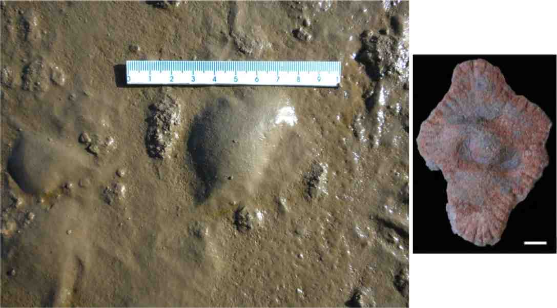
This is a photo of a mature microbial mate with a couple of overturned pieces. Not the cracking in the larger piece around the margin. his may be the cause of the "radial fabric" of the 2.1 GA strucutures. The smaller piece has even more marked cracking and looks similar to this 2.1 Ga specimen below.

This latest evidence strengthens the argument that the 2.1 Ga structures are pyritised microbial mats and not multicellular colonial organisms.
I don't see anything at all like the radial structure exhibited in the fossils in your bubbled mat examples.
ReplyDeleteThe radial structure is caused either by cracking of the pyritised mats during diagenesis or the cracking of the mats prior to pyritisation. Cracks can be seen in the smaller example.
ReplyDeleteThe authors of the original paper have to explain why such a fabric that is essential for the specimens to be a multicellular colonial organism, differs from specimen to specimen, is supposed to be the method of growth at the margins yet does not reach the margins in some, cuts right through a latter pyrite nodule, and is missing altogether in some.