Showing posts with label Cambrian. Show all posts

Saturday, August 13, 2011

Palaeoporn 23

Indiana Nedin and the Temple of Kaili


OK, I'm back, after several months of work and travel, which included losing the top of the Eiffel Tower, and finding it again, avoiding an Ebola E-coli outbreak in Hamburg, and retracing the route of The Italian Job in Torino, it's about time I got back to some real work and started posting on the blog again!

Continuing the tradition of famous fossil sites wot I have visited, the photo above (taken a few years ago now) is of me doing my Indiana Jones impression on a hilltop at the site of the Kaili Formation in Guizhou province, southwest China, some 550 kilometres ENE of Kunming and the Changjiang fauna.

The Kaili formation is basal Middle Cambrian in age (see figure at right) and contains the Kaili Biota, a Changjiang/Burgess Shale type lagerstätte. Given it's age, the biota sits midway in age between the Lower Cambrian Changjiang fauna and the Middle Cambrian Burgess Shale fauna.

As would be expected, the Kaili biota shows significant overlap with both the Chengjiang and Burgess faunas in terms on genera in common. Wikipedia, suggests that of the (quite diverse) 110 genera occurring in the Kaili biota, 30 are shared with the Chengjiang fauna, and 40 are shared with the Burgess fauna.

The Kaili biota is thought to represent an outer-shelf environment and contains large numbers of planktonic trilobites, and eocrinoids, along with a range of soft bodied forms such as neroiids, Wiwaxia and Marrella

However, that's not what I want to talk about. See the village to my right in the photo above (click on the photo to enlarge)? That was our way down from the site. That's what I want to talk about because when we entered the village we were greeted with this:


We had entered a village of the Mountain Miao Minority Peoples of Guizhou, and they laid on a traditional welcome ceremony for us. This started with two local girls in full traditional costume offering food and drink to each person as they entered. The traditional dress included amazing jewelry in solid silver! On this occasion we were offered fish and some lethal alcoholic local brew from a ceremonial horn.

Once inside we were treated to a series of traditional dances, again performed by the girls of the village complete with elaborate gorgeous silver jewelry.


We were not informed that this was going to happen so it was a complete surprise. It was an amazing experience, and was the perfect end to the day.


Further Reading

ZHAO Yuanlong, ZHU Maoyan, Loren E. BABCOCK, YUAN Jinliang, Ronald L. PARSLEY, PENG Jin1, YANG Xinglian1, WANG Yue1 (2005) Kaili Biota: A Taphonomic Window on Diversification of Metazoans from the Basal Middle Cambrian: Guizhou, China. Acta Geologica Sinica; 79(6) 751–765. DOI: 10.1111/j.1755-6724.2005.tb00928.x

Wednesday, February 2, 2011

Palaeoporn 22

Treptichnus pedum


The type section for the Cambrian-Precambrian boundary is at Fortune Head, Burin Peninsula, Newfoundland, as is marked by the first appearance of the trace fossil Treptichnus pedum (the trace fossil formally known as Phycoides pedum).

Treptichnus pedum is a distinctive burrow pattern comprised of a series of lobes set along a central, sometimes curved burrow, thought to represent successive probes into the sediment searching for food. The traces are thought to represent an organism more complex that those of the Ediacaran, but that is not necessarily true, as no actual animal has been found is association with the burrows, so we don't know what made them (although modern priapulid worms make similar burrows).

However, we should not confuse the somewhat complicated pattern of Treptichnus pedum with the first occurrence of well-developed, fairly complex metazoan animals.

While the start of the Cambrian is marked by the first appearance of Treptichnus pedum, this is not the first appearance of trace fossils probably from metazoans. A number of trace fossil types exist in the preceding Ediacaran rocks. In fact they show a neat line in increasing complexity from simple traces early in the Ediacaran to traces as complex as T. pedum towards the end of the Ediacaran.

The earliest traces we have, and the most simple, is Planolites


This is a lowermost Cambrian specimen (picture from NCSE), but it shows you what they are like. The fossil is of simple, cylindrical, unbranched and unlined burrows. They are usually sinuous and undulatory, and often appear as small knobs or discontinuous segments on bedding planes. The small image below is what they typically look like in Ediacaran rocks


Further up in the Ediacaran, more traces appear. This is Helminthopsis


Helminthopsis are much more meandering burrows that appear to represent shallow feeding burrows. the distinctive whorls and loops are considered to represent a different feeding strategy to that of Planolites.

Towards the top of the Ediacaran, things get a bit more complex. As well as Planolites and Helminthopsis, we get the feeding traces of Kimberella.


Here, the feeding traces are marked "R" for radula, and the animal itself is marked "K". A Dickinsonia is also present. "K'" and "D'" mark the resting trace of Kimberella and Dickinsonia, which comprise, of course, another form of trace fossil.

Towards the top of the Ediacaran, things start to get crowded. Not only do we have all the previous traces, but they are joined by Mattaia miettensis.


This specimen comes from the Kessyusa Formation, from Khorbusuonka in northern Siberia. The trace is filled with two bands of sediment, raised on the sides and depressed in the middle, where the bands are separated by a vertical fissure filled with clay. A possible culprit is a pripulid worm such as the Burgess Shale form Louisella pedunculata.

So by the time you get to the Cambrian-Precambrian boundary there were quite a few traces around. Sure in the Cambrian the traces get much more diverse, but they don't start at the boundary, there is a steady increase in trace fossils through the Ediacaran.


Update
Aleksey Nagovitsyn has kindly informed me that the age range for Mattaia miettensis in Palaoeporn 22 is incorrect, and Mattaia miettensis is actually to be found in the Cambrian Tommotian Stage of the Cambrian.

So we need to remove Mattaia miettensis from the list of trace fossils found at the top of the Ediacaran.

Friday, November 5, 2010

Lower Cambrian Sea Anemones from China

Holotype and paratypes of Eolympia pediculata from Han et al. 2010

Yet more exquisitely preserved fossils from the phosphorite deposits in the lowest Cambrian sediments of the Kuanchuanpu Formation, Shaanxi, China. And by "lowest" they really mean lowest! The deposits are only a couple of million years younger that the Cambrian-PreCambrian boundary, which is currently taken as 542 million years ago.

The new fossils have been identified as a possible stem member of the Cnidarian Hexacorallia, suggesting that the diversification of the Cnidaria either occurred very rapidly after the start of the Cambrian, or, more likely (as far as I am concerned), in the Ediacaran.

I don't have much comment to make. The interpretation appears reasonable. The paper is freely available at PLoS (thank you PLoS). I would have liked some larger specimens, but the size is an artifact of the preservation.

There's a nice comparison with some extant polyps from an extant species.

Young polyps from a modern species, from Han et al. 2010

The similarity in form and size is striking. Morphological similarity isn't everything, but it's something!

The authors end with:
The cnidarian diversification might have occurred rather quickly during the early half of the Cambrian or it may be deeply rooted into the Neoproterozoic.
I prefer the latter option, which is a nice intro to Palaeoporn 20!


Han J, Kubota S, Uchida H-o, Stanley GD Jr, Yao X, et al. (2010) Tiny Sea Anemone from the Lower Cambrian of China. PLoS ONE 5(10): e13276. doi:10.1371/journal.pone.0013276

Monday, October 11, 2010

Palaeoporn 19

Another holy grail


Ignore the cute baby buffalo. See that unprepossessing roadside quarry with the people sitting on a pile of rubble (click to enlarge)? Well that rubble is the type section for the Chengjiang fauna.

Yeah! Not so unprepossessing now, is it?!

The Chengjiang fauna occurs in the Maotianshan Shale, a member of the Lower Cambrian Chiungchussu formation. They are found about 5 km northwest of the Fuxian Lake, and 6 km northeast of Chengjiang, Yunnan, China.

The age of the fauna is between 525-520 million years old, significantly older than the Burgess Shale (at 505 million years ago), and the Emu Bay Shale (approx 515 million years old).

The Chengjiang fauna rivals that of the Burgess Shale in preservation and diversity. Although not in difficulty to get to. The Burgess Shale is a 3 hour hike. Here, you can drive right to the outcrop!


In this shot (click to enlarge) you can see the quarry from photo one in the middle distance. There is a bus parked next to it with some people in the road. The hill to the right is Maotian Mount (hence Maotianshan Shale)


In this photo (click to enlarge), I am right at the back in short sleeves and a broad-brimmed brown hat (I'm in the same position in the first photo). I'm there partly because it's in the shade, and partly because there were some nice fossils there (all went to Nanjing University for study).

The depositional environment was delta front prograding eastwards into an open sea. Most of the fossil layers appear to be episodic events onto the marine muds in front of the delta, possible storm induced deposition of clays and fine sand. There's not much evidence of transport, so most organisms were locals and were buried by a series of turbidity flows.


This is a smaller quarry close by (click to enlarge). The dark colour is the fresh colour of the Maotianshan Shale. It is black when fresh, but rapidly oxidises to a tan colour on exposure to air. This is near the top of the Maotianshan Shale. The pick axe handle in the middle of the shot is marking the topmost Maotianshan Shales. The very top of the handle is resting against the first influx of sands, which coarsen upward until they are topped by a large lenticular sandstone at the top of the sequence above my head. This represents the prograding delta as it moves out over the muds of the Maotianshan Shale - just like the sands of the Mississippi Delta are prograding out into the Gulf of Mexico.

Saturday, June 12, 2010

Palaeoporn 15


You spin my head right round, right round, . . .


Carrying on the theme of Palaeoporn 14, namely moulting in trilobites, here’s one to make your head spin.

Literally!

The trilobite at the top of the post is a big Redlichia takooensis (around 12-14 cm in length), common in the Lower Cambrian Emu Bay Shale, except that it's head is on backwards and inside out!

First a little background. The trilobite exoskeleton is rigid, and so to grow they need to shed this outer covering, expand, and mineralize another exoskeleton around the expanded body (crabs do this today, with soft crabs – those that have shed their exoskeleton and are awaiting the new exoskeleton to harden - prized as bait.)

Since the exoskeleton is rigid, there needs to be an exit strategy so that the body can get out of the old exoskeleton. This is usually achieved in trilobites by having lines of weakness – or sutures – at strategic places on the body, which preferentially break. These are usually placed on the head and around the eyes, and separate the central part of the head - or fixigena - from the outer part of the head - or librigena - which is also called the free cheeks 'cos they represent the cheeks of the head and they get freed up during moulting.

When the trilobite starts moulting, usually the suture lines break, the exoskeleton around the head separates into fragments (fixigena and librigena), allowing the body to exit through the head, leaving the exoskeleton intact, and the head fragments to fall back into place.

Usually.

Sometimes it doesn’t go to plan.

The free cheeks can be displaced, but that is usually the trilobite being careless on the way out. But sometimes things go wrong.

For comparison here is a proper R. takooensis (right, trilobite length 5 cm without spine) with its head on straight, fixigena and librigena all facing front and orientated correctly, even if the free cheeks are slightly displaced indicating that it is a moult.

So what happened to our backward friend up front?

Well, I'm pretty sure it wasn't born that way (no offense Jake), so it looks like a moulting accident.

Also the librigena isn't.

Liberated that is.

Although we don't have the whole body, the portion of the head outside of the eye (the librigena) is still in place indicating that the suture did not split.

What probably happened was that once the facial sutures failed to split, the exoskeleton broke behind the head. The animal then exited the old exoskeleton, pushing the head exoskeleton into the vertical and then beyond, which forced the head exoskeleton upside down and the front margin to point backwards.

In other words, imagine the head is an upside down bowl as in the diagram below, where F = front of the head, and B = back of the head.


Instead of splitting along the sutures, the whole head exoskeleton comes detached from the body exoskeleton. The trilobite then pushes its way out by forcing the head exoskeleton to tip 90 degrees onto the front margin, and then 180 degrees to lie upside down, with the front margin now pointing backwards and exposing the internal surface of the head exoskeleton.

The trilobite then escapes the old exoskeleton and is free to go.

Scary stuff perhaps, but not the worst example.

Next time - "Ultimate Moulting - when moulting REALLY goes bad"

Thursday, June 3, 2010

Squid Wannabes in the Cambrian

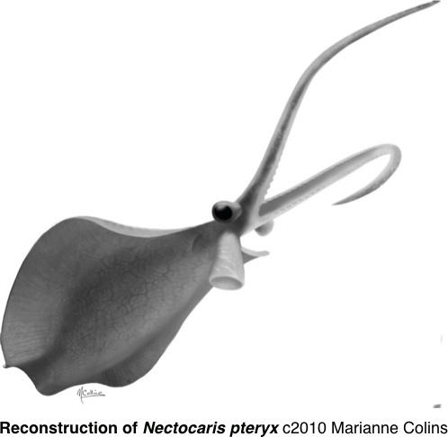
ResearchBlogging.org Another problematic Cambrian form finds a home. Once more the Burgess Shale comes up trumps, with the work of Martin Smith and Jean-Bernard Caron from the University of Toronto/Royal Ontario Museum shedding new light on Cambrian critters and the evolutionary things they get up to.

Ok. This is neat, and a group that occurs in the Burgess Shale, the Emu Bay Shale, and Chengjiang. The Burgess Shale form Nectocaris pteryx, and the closely related forms Vetustovermis from the Lower Cambrian Emu Bay Shale,

and the now not-synonymous Petalilium from the Lower Cambrian Chengjiang fauna, has been re-interpreted as a stem group cephalopod.

The arguments in favour of the forms being stem group cephalopods is persuasive (stem group forms lack one or more features characteristic of the last common ancester of the crown group).

The forms have a number of characters that link them with molluscs, and closely with cephalopods. These include the presence of tentacles - albeit only one pair, an axial cavity containing gills (possibly homologous with the mantle cavity of crown group cephalopods), and a funnel

They are also rare – ninety-odd specimens from the Burgess Shale may seem a lot, but it isn’t really. The Emu Bay Shale form Vetustovermis is very rare. I didn’t find one decent specimen when I worked on the deposit. But rare is good if you are trying to push the mollusc line, because molluscs don’t moult. Arthropods do. And moulds can fossilise. In effect, this is like leaving numerous photocopies of yourself in the fossil record. One arthropod can leave numerous fossils behind. Molluscs can’t. So we would expect them to be rarer than arthropods, as is the case here.

The eyes are interesting. They are preserved differently that other eyes in the Burgess Shale. Usually, eyes are preserved as a carbon film coated by clay minerals. This is similar to body preservation and is taken to indicate that the eyes were compound (made of calcite crystals) and thus robust enough to preserve the same way as the body. In Nectocaris (as in the similar Chengjiang form Petalilium) the eyes are preserved as a carbon film that covers a thick layer of muscovite crystals. This is interpreted by the authors as indicating that the eyes were hollow in life, similar to cephalopod (and our) eyes today, rather than the compound eyes of arthropods.

Another nice feature is the serial repeated pairs of gills. Modern cephalopods have one gill, or set of gills, but the sequence of repeated pairs of gills in Nectocaris (and in Petalilium, and Vetustovermis) is exposing its common ancestry with segmented forms. In other words the common ancester of molluscs and arthropods was a metamerically segmented form (a form with a series of similar segments, like a trilobite or worm). Nectocaris, with its sequence of repeated pairs of gills, is therefore, a neat intermediary between the metamerically segmented ancestral form and the derived, more modern forms that have lost the segmentation. In mean, if you'd have asked a palaeontologist what a stem group cephalopod would look like, the answer would have been paired gills all the way down!

This group appears to lack a horny beak, a radula, a shell, and at least eight tentacles, which is why they are considered stem group forms. The last common ancester of the cephalopods is considered to have had all of these.

I have a few issues however.

First, have to say I’m not a fan of the paper’s title, Primative soft-bodied cephalopods from the Cambrian. “Primitive”! Oh dear, I had though we had stopped using that term – Early perhaps). And they are not strictly cephalopods (they are however, Conchifera). So, "Early Conchiferids from the Cambrian" perhaps (a bit dry I’d admit), or my personal choice, "Squid wannabes from the Cambrian".

Second, I am not a fan of the reconstruction either. Not the drawing itself – I’m a big fan of Marianne Collins’ work – but of the way it hovers with the funnel aimed downward like a Harrier Jump Jet or, as in Nature News and Views, the rocket underneath the Space Shuttle. Ugh!

It’s very unlikely that the funnel would have been used like that. One of the specimens has it in that position (figure “f” in first image) but it is unlikely to represent the life position. Burgess Shale fossils are found in all orientations, and numerous other specimens of Nectocaris have the funnel in various orientations. I think that figure “f” has a bad case of flacid funnel, probably post mortem.

The funnel was probably used to move forwards and backwards, but also maybe to blow fine sediment away from shallowly buried prey, or even blow them so that they tumbled which disorientated them, make them easier to catch. But what prey did they hunt? This is especially interesting given the jaws, or rather the lack of them! Which brings us to . . .

Third, where’s the jaws? It looks like they are absent in Nectocaris. This is strange, as the presence of teeth or radulas are well established in the Mollusca by the Middle Cambrian Burgess Shale time. Modern cephalapods have a beak, but the radula is reduced in octopus, and is absent (or extremely reduced) in Spirula, the Ram’s Horn Squid.

The authors say that the absence could be due to it not being preserved or that it is too small to preserve. I’m not buying that it didn’t preserve. Hard parts of other organisms preserve just fine in the Burgess Shale. But it could be that they were very small. Spirula is a small (around 4 cm) deep water squid that either has a very small or non-existant radula. Spirula feeds on plankton, so it could be that Nectocaris also fed on tiny plankton as well.

There is some vague feature which the authors claim could be mouth parts. If so it would suggest a diet of soft bodies organisms or very small organisms such as plankton.

So three Cambrian forms tidied up quite nicely, and a neat transitional form (gasp!) as well! Cambrian squid wannabes with a hangover from their metamerically segmented ancestry.


Smith, M., & Caron, J. (2010). Primitive soft-bodied cephalopods from the Cambrian Nature, 465 (7297), 469-472 DOI: 10.1038/nature09068

Smith, M., & Caron, J. (2010a). Primitive soft-bodied cephalopods from the Cambrian: Supplimentary Information Nature, 465 (7297), 469-472 DOI: 10.1038/nature09068

Chen, Jun-yuan; Huang, Di-ying; Bottjer, David J. (2005). "An Early Cambrian problematic fossil: Vetustovermis and its possible affinities.". Proceedings of the Royal Society, Part B 272 (1576): 2003–2007. doi:10.1098/rspb.2005.3159.

Sunday, May 30, 2010

Cambrian Critters in the Ordovician 2

ResearchBlogging.orgDuring the 46-million-year Ordovician Period (489–443 mya), a phenomenal array of adaptive radiations of "Paleozoic- and Modern-type" biotas appeared in marine habitats, the first animals walked on land, and the plants appeared.

This Great Ordovician Biodiversification Event represents a tripling of diversity at the family level, and a quadrupling at the genus level, from those in the Cambrian.

The number of families reached by the end of the Ordovician remained fairly constant (except for a number of mass extinction events) for almost 200 million years. Articulate brachiopods, conodonts, graptolites molluscs, crinoids, and trilobite groups all became established of diversified greatly during this time.


A major question is what happened to the Cambrian faunas - as typified by the Burgess Shale, the 'soft-bodied', or poorly mineralised, organisms that are found in numerous Cambrian deposits. They are by-and-large absent from the Ordovician.

We do have a large number of Cambrian sites with exceptional preservation (the Bugress Shale being the ‘type’ example), but from the Ordovician, not so much. In fact very few. In fact, bugger all!

What Ordovician exceptional preservation we have comes from the middle and late Ordovician, the Beecher’s Trilobite Bed, New York, the Soom Shale, South Africa, Winneshiek, Iowa, and a couple of localities in Manitoba, Canada.

All localities represent restricted faunas from extreme environments (low oxygen) and not examples of more diverse, open marine environments. (Yes, the Burgess Shale may well represent a low oxygen environment, but the biota it contains represents a diverse open marine environment.)

No examples of exceptional preservation have been found from the early Ordovician.

Given this extreme lack of exceptional preservation, our understanding of this great biodiversification event is almost entirely based on the shelly fossil record. What we get, is a spectacular rise in the shelly fossil record.

This leads to the obvious question. What happened to the Cambrian critters?

There are two main theories to explain that wholescale change in faunas. One is that the Cambrian forms were replaced by Ordovician forms. The other is that the exceptional preservation we see in Cambrian deposits simply does not occur in the Ordovician, and so the missing forms are due to a taphanomic, or preservational, bias. So which is it? replacement of preservational?

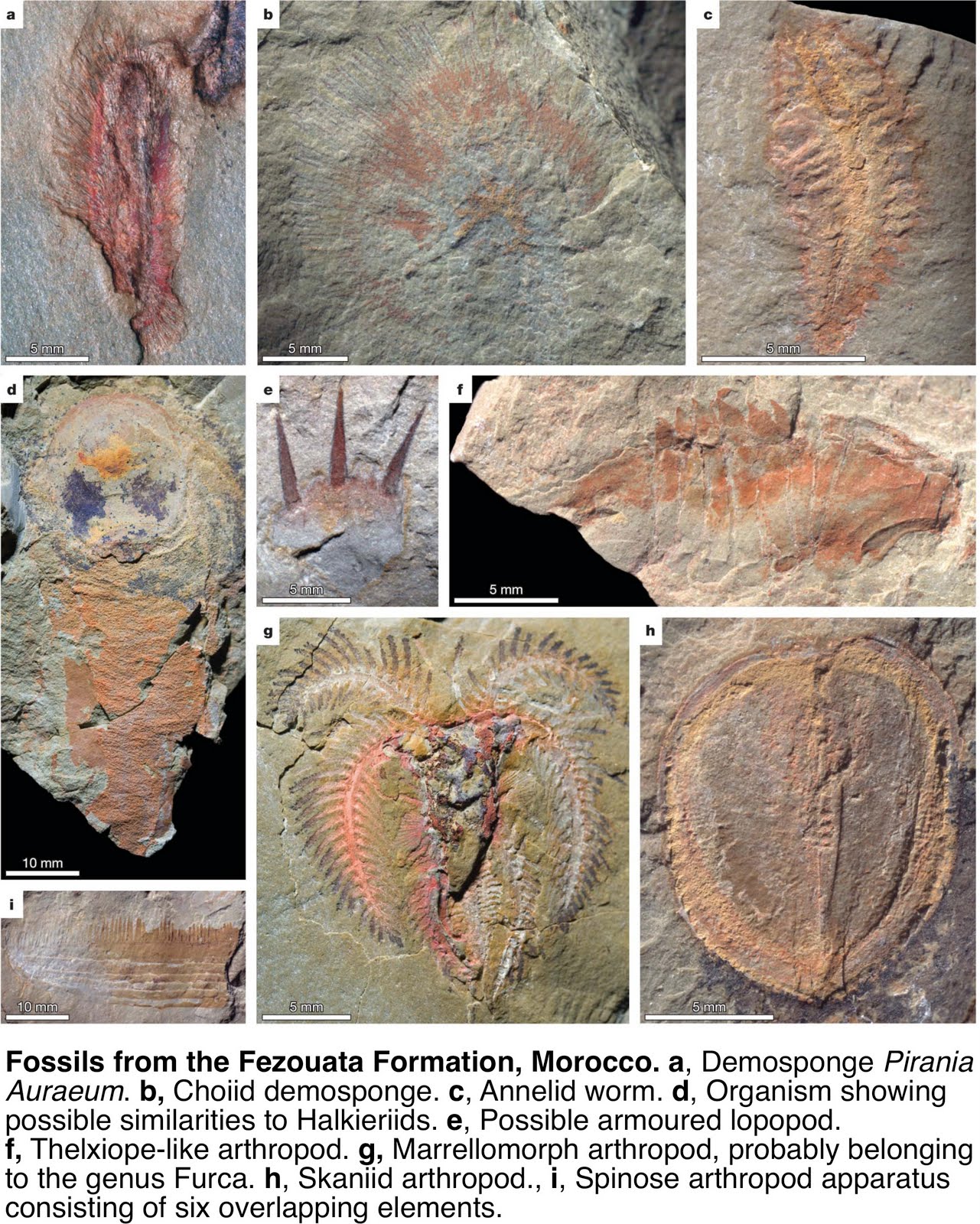
Introducing the Fezouata biota from the lower Ordovician of Morocco.

This represents the first site of exceptional preservation from the lower Ordovician, and (if that wasn’t important enough) the first from a ‘normal’ open marine environment.

Over 50 different taxa have been collected so far indicating an open marine, deep water assemblage that has been trapped beneath, or brought in by, storm deposits. Limited bioturbation indicates low oxygen which would account for the exceptional preservation. The fauna is a mix of typical Ordovician forms and typical Cambrian Burgess Shale-types, although there are similarities with the Chengjiang fauna from China, both in terms of fossils and depositional environment.


One cool find is a xiphosurid (a horseshoe crab - everyone's favourite chelicerate). The horseshoe crab fossils are the oldest yet found, suggesting that their roots may dip into the Cambrian. What is really cool is that one of the two species of horseshoe crab has a fully segmented opisthosoma (it is either partially or fully fused in other forms), suggesting evolution from a segmented ancester. The other has a fully fused preabdomen (quite a derived feature) and is similar to existing forms (though not the same.)

This has major implications for the transition from Cambrian fauna to the Palaeozoic fauna represented by Ordovician forms. Namely that the Cambrian critters hung on for a considerable time after the end of the Cambrian and intermingled with Ordovician forms. So there was no wholesale replacement at the end of the Cambrian. Both sets mixed.

However it is interesting to note that this deep water deposit was close to the South Pole during lower Ordovician times. The deposits represent a deep, cold water environment. Cambrian deposits with exceptional preservation, by contrast, were primarily from low latitudes, near the paleoequator, and from shallow water.

It may be that the Fezouata biota represents a refuge for the Cambrian forms - much like modern brachiopods are excluded from tropical areas and are mainly found in temperate and cold deep water today.

So this biota shows that Cambrian forms persisted well into the Ordovician, but a shallow water site of exceptional preservation is needed to see if the Cambrian critters were mixing it with the Ordovician upstarts, or were just hanging on along the margins.

Thanks to Paul H.

Van Roy, P., Orr, P., Botting, J., Muir, L., Vinther, J., Lefebvre, B., Hariri, K., & Briggs, D. (2010). Ordovician faunas of Burgess Shale type Nature, 465 (7295), 215-218 DOI: 10.1038/nature09038

Saturday, May 15, 2010

Cambrian Critters in the Ordovician

There's a paper in Nature on a new Ordovician site with a range of critters resembling Cambrian fossils.

I am waiting to get my hands on a copy of the paper to comment, but until then Brian Switek has it covered here.

One thing though. The deposit is apparently from deep water. This may well be an example of critters being forced out of the near shore by competition and hanging on in a deep water 'refuge'.

More once I get the paper.

Monday, April 26, 2010

Palaeoporn 14

Ménage à trois!

These are Estangia bilobata trilobites from the Lower Cambrian Emu Bay Shale on Kangaroo Island, South Australia. Estangia is the most common fossil found in the Emu Bay Shale. However, these came from the outcrop of Emu Bay Shale at Emu Bay, and not from the more famous site further along the coast that contains exceptionally preserved fossils such as Anomalocaris and Myoscolex. (The Emu Bay Shale outcrops at two locations on Kangaroo Island)

These little critters show the difference between the two depositional sites. The site with exceptional preservation shows evidence - palaeontological (whole specimens, low diversity) sedimentological (fine grained sediments), and chemical (evidence of reducing environment-enriched trace elements) - of a low energy, low oxygen environment conducive to exceptional preservation.

On the other hand at Emu Bay the site shows evidence of a higher energy, higher oxygen depositional environment. This is because the sediments show more interbedded sands and silts (coarser grained therefore higher energy), oxidating environment-enriched trace elements, and the fossils do not show exceptional preservation, and are fragmented.

In the example above, the fossils assemblage comprises the heads of three Estangia trilobites (the lower one is both turned over and spun through 180 degrees). The heads are not complete. The sides of the head - the librigena (or the free cheeks) are missing. This shows that the heads represent molts.

Trilobites are arthropods and so have to molt the outer exoskeleton in order to grow. To do this, they have special lines of weakness in the exoskeleton called sutures. When the trilobite molts, these suture lines break apart, allowing the trilobite to leave the exoskeleton. These sutures are particularly obvious on the head where they run from the margin down to the eye, around the eye and then back out to either the side margin or the back margin.

In this case the suture lines are opisthoparian,
as they run from the eye to the back of the head rather than out to the side They run along the eye so that the eyes will be the first thing to break out of the old exoskeleton - allowing the trilobite to keep its vision while the molting process continues. This results in the librigena breaking away from the head. Often the molting process breaks the attachment between the head and the rest of the body, resulting in the head becoming detached from the rest of the body.

Since these fossils comprise the cranidium only (head minus the librigena), this indicates that they are molts and that they have been sorted by currents that have separated them from the body and librigena.

Compare this with another Emu Bay Shale Estangia, this time from the site of exceptional preservation (right).

Here the head and body is present. However, it is still a molt because the librigena have been freed from the head. In this specimen, the right librigena (outlined) is still associated with the body but has moved some distance away, and is both turned over and spun through 180 degrees so the the spine (that normally points backwards) now points forwards.

So the three in the top image represent a disarticulated random grouping, and have not been caught in flagrante delicto. So move along . . . nothing to see here . ..

Diagram credit:
Trilobite Facial Sutures

Sunday, May 24, 2009

Palaeoporn 13


Look up there! Is it a brachiopod? Is it an annelid? Is it a mollusc?

Umm  . . . actually, its a bit of each really.

This is a wiwaxiid from the Emu Bay Shale. Unfortunately undescribed, but closely linked to Wiwaxia and the halkieriids. It would be one of the oldest examples of a wiwaxiid, as the others are from the Middle Cambrian Burgess Shale and comparable deposits.

The beast was bilaterally symmetrical, oval in shape, and covered with short scales - called sclerites. Also present were a number of rows of larger spines which protruded upwards in a probable defensive array (see reconstruction at right)

Wiwaxiids had a flat foot-like underside. Little is known of the internal anatomy.

Wiwaxiids have been classified as molluscs, annelids and stem group annelids (a group closely related to annelids). The spines have been compared to the eltrya, or scales, of polychaete worms, and even bristles of molluscs, annelids and brachiopods, and halkieriids, of course, have little brachiopod shell caps!

One feature found in wiwaxiids is a radula-like feeding bar. So I looked to see if my specimens had a radula-like feeding bar. I've switched to black and white photos for higher resolution. A bar structure, formed of calcium phosphate, was found towards the front of the specimen. After preparing out (lower image) half the bar remained and the other half (the distal, or outer, part of the bar is removed, leaving a mold of the lower surface of the bar. in this mold can be seen several depressions along the 'upper' margin, represented by shadow (the light is coming from the top right of the image). These are 'teeth' which would have protruded from the lower margin of the bar (the bar is approx. 5 mm long.)

So, spines, radula-like feeding bar, seems like a Lower Cambrian wiwaxiid!

Wednesday, April 15, 2009

The World's Oldest Poo

You can learn a lot from poo.

Fossil poo, or coprolites, can provide valuable information on the size and feeding habits of the organism that produced them.

Large Cambrian coprolites are rare, and tend to be circular, but one found in the Lower Cambrian Emu Bay Shale was different.

Before going further, you should look at this short TV story that was done last year. It puts the poo in context, and the poo has a small cameo.

Keeping with the theme of using black and white photography to show critical features that may not be apparent in colour (in Palaeoporn 12), I have a black and white image of the poo. But before that I need to show you some features of Redlichia highlighted in the following image (click to enlarge).


Now pay attention as there'll be a test later. The left hand image shows a complete Redlichia. The feature in the blue box is a thorasic spine. Redlichia had a number of these attached to a number of axial rings, which comprises the central ridge. The central image shows a close up of the tip of the segment in the yellow box, showing the tip of a depression in the segment, or pluron, called the plural furrow. The right hand image shows the head region of a large Redlichia. Around the whole of the outer margin of the head is a thick zone with terrace ridges (in the red boxes) that are common in Redlichia. (Incidentally the structure coming off the head at one of the red boxes, is an antenna).

Ok, now for the image of the poo (click to enlarge).

The lower image has been augmented to delineate the poo and to highlight certain features.

Firstly the size. it's 43 mm in length and has a maximum width of 28 mm. It is formed of two 'layers' an upper diffuse layer, probably representing a fluid phase that has expanded outward. The lower layer is coarse particulate layer is crammed with trilobite fragments. If you compare the boxed areas of the poo with the three part image above (colour coded for your convenience), you should be able to make out just which parts of the ex-trilobite are represented here. The whole of the lower part of the poo is trilobite hash!

The poo tells us what was being eaten. The thorasic spine, plural tips and terrace lines present identify the remains as that of a Redlichia.

The poo tells us the size of what was being eaten - about 4 cm in length.

The poo tells us how it was being eaten. The fragments are broken. They are not crushed. They are not nibbled. They are not gnawed. They are not bitten. This means that the trilobite was broken up and not bitten (see CSI-Cambrian).

The poo tells us the size of what was doing the eating. Assuming the trilobite hash layer represents the true original thickness of the poo is 15 mm. This gives us a terminal alimentary tract of 15 mm, which roughly correlates with a 75 cm body size (from measurements done of other Cambrian fossils).

The poo tells us what was doing the eating. The broken up fragments and the size of the organism suggests Anomalocaris was the perpetrator.

So this one specimen tell us that two particular species were present, which was the predator, which the prey, and the method of predation.

Not bad for a piece of crap!

Wednesday, April 8, 2009

Show us some leg

Now that I have your attention, I'd like to talk legs. No, not the Blahnik-clad ones opposite (a homage to the Goddess), but arthropod legs (sorry).

When I talked about the new reconstruction of the anomalocarid Hurdia from the Burgess Shale, I mentioned how it supported a particular interpretation of the evolution of the arthropod limb. Now I'd like to go into a bit more detail on this because it raises some interesting questions about what was going on down in the Cambrian, especially concerning arthropods.

Now the first thing to remember, is that in the Cambrian there were lots of almost-arthropod groups scurrying around and messing up a nice, neat, columbiformes-holed phylogenetic classification, in which arthropods were real arthropods, and ancestral and derived features knew their place. Biology is messy, especially in the Cambrian, deal with it.

Basically there were two groups - the Euarthropods, and everyone else. The Euarthropods are what we would consider to be the 'true' arthropods, with all the features we have decided can be used to classify them as arthopods (except where they have been secondarily lost, for what were probably very good reasons at the time) (we could go into symplesiomorphies, apomorphies, and all the other morphies, but frankly, that way lies madness, otherwise known as cladistics).

The groups outside the Euarthropods, such as the Megacheria, the Radiodonia, can either be considered as extremely interesting groups in their own right, with many of the features of 'true' arthropods, and have much to tell us about evolution in general, and arthropod evolution in particular - or as pathetic arthropod wannabes. Almost-arthropods that failed to make the grade.

My position is, I think, clear, Anomalocaris RULES.

Anyhow, back to legs (no, not the Blahniks - concentrate). Your bog standard arthropod leg is biramous. This means that it has two main components, the walking leg (the endopodite) and the frilly bit, which is the gill (the exopodite). (see diagram at right).


It was thought that the biramous limb is an ancestral condition, and was formed by the fusion of a separate walking limb with a frilly bit. So when we look at the other groups in the Cambrian what we see are separate limbs. Hurdia is a good example. The image at right is the gill structure found in Hurdia. A nice big gill-like structure, but no walking limbs. So Hurdia supports the idea that in groups ancestral to Euarthropods, the walking limb and gill were separate structures. All well and good.

However, work by Wolff and Scholtz1 has thrown some doubt on this neat idea.

They looked at the formation of both biramous and uniramous limbs in the crustacean Orchestia, which has both types.

They found that the biramous limb was formed by a secondary subdivision of the growth zone of the main limb axis to produce the endopodite and exopodite. But, the uniramous limb was not formed by the loss of the exopodite but by suppression of the split into exopod and endopod.

They argue that, if the biramous limb was formed by fusing two separate structures, the subsequent production of a uniramous limb would occur through the loss the exopodite. Whereas here, the exopodite never forms, and so the production of the euarthropod biramous limb is through splitting of the main limb axis during development, and not by the fusion of two separate structures.

Thus, the euarthropod biramous limb has developed from within the euarthropods, rather than be an ancestral feature.

The fossil record supports separate limbs for the pre-euarthropods, and I don't think the fusion theory is completely discredited yet. It could be that the fusion of both structures has linked their development to a large extent. But the findings are really interesting, and shows that we are still learning about early evolution during the Cambrian.

Biology! It's messy. It's complex. It's a harsh mistress. It's a scientific discipline in Blahnik boots.

1Carsten Wolff and Gerhard Scholtz (2008) The clonal composition of biramous and uniramous arthropod limbs. Proceedings of the Royal Society, B. pp 1-6. doi:10.1098/rspb.2007.1327

Tuesday, March 10, 2009

Palaeoporn 11 - again


OK, back by huge public demand . . . err of one. Palaeoporn 11 is back with mark-ups.

The photo is of a portion of an Anomalocaris appendage, and shows the typical preservation style of fossils in the Emu Bay Shale.

In the photo (click to enlarge) there are three segments (S) and two segment boundaries (SB) the segment boundary is doubled because the original appendage was roughly circular, and when you flatten a tube, you don't get the appendage boundary matching up when squashed.

The specimen has been re-crystallised during an episode of overthrusting. The surface of the fossil is normal calcite, but in the centre the calicite is in the a fibrous form (FC). The matrix where the fossil was, is smooth (M2), the normal matrix is M1.

There is no phosphatisation of muscle tissue, and so this is probably a moult. But that's another story . . .

Sunday, March 8, 2009

Palaeoporn 11


A slightly different Palaeoporn this time, as there's a bit of a theme this week, on preservation. A bit later I'll be discussing how to fossilise muscle tissue. This, and other specimens helped in coming up with the process.

This is a fragment of an Anomalocaris appendage from the Cambrian Emu Bay Shale. The interesting thing is the preservation. The outer layer preserves the impression of the appendage, together with segment boundaries. Inside, is a layer of fibrous calcite crystals. The specimen has been re-crystallised after a metamorphic event which included overthrusting. The crystals are, in fact aligned to the stress field, as they 'lean' towards the north - the thrust direction was south to north.

Tuesday, February 17, 2009

Palaeoporn 10


This is Isoxys communis a phyllocarid crustacean from the Lower Cambrian Emu Bay Shale, in South Australia. (click on the image to enlarge)

Isoxys is an arthropod with a curved bi-valved carapace, inside which the organism lives. In this case, all the organism lived within the cavity created by the carapace, except for the tips of the appendages, and, visible in the specimen above, eyes. This is the first occurrence of eyes in I communis, and allowed the correct orientation of the organism to be established. I communis has two spines along the top of the organism, pointing forwards and backwards. The backwards facing spine is longer that the forward facing spine, but the original reconstruction of I communis had the long spine facing forwards.

The specimen above shows two stalked eyes, and most of the organism, with the posterior missing. If you check the enlarged image, and follow the margin of the bi-valved carapace from behind the eyes downwards and around the bottom of the animal, the carapace ends at a fracture line. However a thin pink line in the matrix continues on backwards. This is the other valve of the carapace, showing the sediment filling most of the cavity between the bi-valves.

The forward spine on the specimen with eyes above is almost the correct size, just the tip is missing. The larger specimen lying directly beneath it is showing the posterior spine, it's much longer

Isoxys is quite common in the Emu Bay Shale fauna (there are parts of 4 specimens in the sample above), making up 13% of the fossils. The genus is also found in the Burgess Shale, and in the Chengjiang fauna in China, but they are whimps compared with I communis, which is up to 4 times bigger. Go Aussie!

Sunday, January 11, 2009

Myoscolex - a Lower Cambrian Opabinid


This is Myoscolex ateles (click on the images to enlarge), a common element of the Emu Bay Shale faunal assemblage – which is a restricted fauna of only some dozen species (this compares with over 150 for the Burgess Shale). Why the Emu Bay Shale is such a restricted fauna will be covered in a future post on fossilisation.

The preservation of Myoscolex is striking. The grey material is calcium phosphate (apatite) and the red material is calcium carbonate (calcite) – in this case the calcite is just a light dusting over the apatite. The material preserved is muscle tissue – one of the earliest examples of phosphatised muscle tissue known. The colours actually pick out the two sets of muscles in the organism. The grey apatite highlights the longitudinal (along the body) muscle bands, while the red area highlights the dorso-ventral (top to bottom) muscle bands.

One feature not present in all specimens are rod-like structures. In the original description of Myoscolex from 1979, these rod-like structures were interpreted as paripodial setae, and the animal classed as a polychaete worm, although it was admitted that the setae were different from any yet known in polychaete worms.

A number of factors, including the collection of more specimens with new information, and a better understanding of soft tissue fossilisation, resulted in a re-evaluation of Myoscolex as part of my work on the Emu Bay Shale.

It was found that Myoscolex is actually quite common, around 15% of all fossils found (trilobites were the most common at just over 50%), and that pretty much all the Myoscolex specimens found were in the same orientation - laterally flattened and straight (as in the first image). This was unusual in polychaetes, who tend to be found dorso-ventrally (top to bottom) flattened, and exhibit a greater range of flexibility.

A close analysis of the rod-like structures showed that they were paired, and extend out from the body itself. Not only that but they were preserved at different levels on the fossil. Check out the second image showing the rods. You can see the pairs, but one rod is missing, with only the imprint of where it was. This indicated that in the fossil, the pair of rods were lying at slightly different levels in the rock. In this instance, the missing rod was slightly higher in the rock and has separated with the upper part of the fossil when the rock split. The fact that the rods extended some distance from the body (as represented by the muscle tissue), and that they are not level, suggests that they have something to do with endoskeletal sternites, or the lower portion of the body segment. Which means that the animal must have come to rest on it's side and then been flattened so that the sternites folded up into a "U" shape with the lateral (outer) edges coming to rest close together like the top of the "U".

Except worms do not have sternites. Hmmm.

Then there was a breakthrough with this single specimen, which showed beautiful appendages.


And they don't look like appendages any worm would have. They are paired and extend a considerable distance from the body of the animal. Plus the appendages differ in their orientation along the body, indicating that they were flexible. This find also explained why almost all the fossils were found in lateral aspect. The appendages would have come together under the animal upon death or burial, with lying on the side the most stable position. The appendages also showed a variation in attitude along the body, indicating a degree of independent flexibility.

This specimen also showed a portion of the tail, which pointed upwards.

These findings alone were enough to re-evaluate the animal, but a specimen with a head turned up, and things got weird.


Nice eyes (E), shame about the number! Wait a minute, 3 eyes?! 3?! Yeah, and they aren't the same either. The two at the top in the specimen are sub-circular, but the one on the bottom is large elongate structure, which could be one long eye, or two or three that are overlapping ue to twisting of the head. The eyes are also preserved differently - as featureless films of fibrous calcite, not apatite, which indicated that there is no muscle tissue involved (as expected in eyes).

Not only that but there is a faint structure running backwards along the bottom of the head and start of the body (P). It's not well preserved, but is formed of apatite which means that it had contained muscle tissue, and is interpreted as a proboscis. Another specimen showing a possible proboscis is shown at right.

So what type of organism has more than two eyes, a proboscis, flappy paddle appendages, and an upwards pointing tail?

Well, the obvious candidate is Opabinia from the Middle Cambrian Burgess Shale. A check of the numerous reconstructions of Opabinia show that the same suite of characters are present, even the change in orientation of the paddle appendages along the body.

So Myoscolex has been reclassified as an arthropod of similar affinities to Opabinia. So Myoscolex is an Opabinid, or maybe Opabinia is a Myosolexid, as Myoscolex is older than Opabinia ;-)