OK, I'm back, after several months of work and travel, which included losing the top of the Eiffel Tower, and finding it again, avoiding an Ebola E-coli outbreak in Hamburg, and retracing the route of The Italian Job in Torino, it's about time I got back to some real work and started posting on the blog again!
Continuing the tradition of famousfossilsites wot I have visited, the photo above (taken a few years ago now) is of me doing my Indiana Jones impression on a hilltop at the site of the Kaili Formation in Guizhou province, southwest China, some 550 kilometres ENE of Kunming and the Changjiang fauna.
The Kaili formation is basal Middle Cambrian in age (see figure at right) and contains the Kaili Biota, a Changjiang/Burgess Shale type lagerstätte. Given it's age, the biota sits midway in age between the Lower Cambrian Changjiang fauna and the Middle Cambrian Burgess Shale fauna.
As would be expected, the Kaili biota shows significant overlap with both the Chengjiang and Burgess faunas in terms on genera in common. Wikipedia, suggests that of the (quite diverse) 110 genera occurring in the Kaili biota, 30 are shared with the Chengjiang fauna, and 40 are shared with the Burgess fauna.
The Kaili biota is thought to represent an outer-shelf environment and contains large numbers of planktonic trilobites, and eocrinoids, along with a range of soft bodied forms such as neroiids, Wiwaxia and Marrella
However, that's not what I want to talk about. See the village to my right in the photo above (click on the photo to enlarge)? That was our way down from the site. That's what I want to talk about because when we entered the village we were greeted with this:
We had entered a village of the Mountain Miao Minority Peoples of Guizhou, and they laid on a traditional welcome ceremony for us. This started with two local girls in full traditional costume offering food and drink to each person as they entered. The traditional dress included amazing jewelry in solid silver! On this occasion we were offered fish and some lethal alcoholic local brew from a ceremonial horn.
Once inside we were treated to a series of traditional dances, again performed by the girls of the village complete with elaborate gorgeous silver jewelry.
We were not informed that this was going to happen so it was a complete surprise. It was an amazing experience, and was the perfect end to the day.
Further Reading
ZHAO Yuanlong, ZHU Maoyan, Loren E. BABCOCK, YUAN Jinliang, Ronald L. PARSLEY, PENG Jin1, YANG Xinglian1, WANG Yue1 (2005) Kaili Biota: A Taphonomic Window on Diversification of Metazoans from the Basal Middle Cambrian: Guizhou, China. Acta Geologica Sinica; 79(6) 751–765. DOI: 10.1111/j.1755-6724.2005.tb00928.x
The type section for the Cambrian-Precambrian boundary is at Fortune Head, Burin Peninsula, Newfoundland, as is marked by the first appearance of the trace fossil Treptichnus pedum (the trace fossil formally known as Phycoides pedum).
Treptichnus pedum is a distinctive burrow pattern comprised of a series of lobes set along a central, sometimes curved burrow, thought to represent successive probes into the sediment searching for food. The traces are thought to represent an organism more complex that those of the Ediacaran, but that is not necessarily true, as no actual animal has been found is association with the burrows, so we don't know what made them (although modern priapulid worms make similar burrows).
However, we should not confuse the somewhat complicated pattern of Treptichnus pedum with the first occurrence of well-developed, fairly complex metazoan animals.
While the start of the Cambrian is marked by the first appearance of Treptichnus pedum, this is not the first appearance of trace fossils probably from metazoans. A number of trace fossil types exist in the preceding Ediacaran rocks. In fact they show a neat line in increasing complexity from simple traces early in the Ediacaran to traces as complex as T. pedum towards the end of the Ediacaran.
The earliest traces we have, and the most simple, is Planolites
This is a lowermost Cambrian specimen (picture from NCSE), but it shows you what they are like. The fossil is of simple, cylindrical, unbranched and unlined burrows. They are usually sinuous and undulatory, and often appear as small knobs or discontinuous segments on bedding planes. The small image below is what they typically look like in Ediacaran rocks
Further up in the Ediacaran, more traces appear. This is Helminthopsis
Helminthopsis are much more meandering burrows that appear to represent shallow feeding burrows. the distinctive whorls and loops are considered to represent a different feeding strategy to that of Planolites.
Towards the top of the Ediacaran, things get a bit more complex. As well as Planolites and Helminthopsis, we get the feeding traces of Kimberella.
Here, the feeding traces are marked "R" for radula, and the animal itself is marked "K". A Dickinsonia is also present. "K'" and "D'" mark the resting trace of Kimberella and Dickinsonia, which comprise, of course, another form of trace fossil.
Towards the top of the Ediacaran, things start to get crowded. Not only do we have all the previous traces, but they are joined by Mattaia miettensis.
This specimen comes from the Kessyusa Formation, from Khorbusuonka in northern Siberia. The trace is filled with two bands of sediment, raised on the sides and depressed in the middle, where the bands are separated by a vertical fissure filled with clay. A possible culprit is a pripulid worm such as the Burgess Shale form Louisella pedunculata.
So by the time you get to the Cambrian-Precambrian boundary there were quite a few traces around. Sure in the Cambrian the traces get much more diverse, but they don't start at the boundary, there is a steady increase in trace fossils through the Ediacaran.
Update
Aleksey Nagovitsyn has kindly informed me that the age range for Mattaia miettensis in Palaoeporn 22 is incorrect, and Mattaia miettensis is actually to be found in the Cambrian Tommotian Stage of the Cambrian.
So we need to remove Mattaia miettensis from the list of trace fossils found at the top of the Ediacaran.
Frondose (frond-bearing) forms are the most complex and interesting of the Ediacaran fossils, but rare. This is because most of the organism lives above the sea floor, to which they are anchored by a round holdfast, and so are not preserved. A number of frondose forms have been found, but Pambikalbae is different.
Pambikalbae hasenohrae(a) complete block showing several specimens (upper right and lower left). Slab 45 cms. (b) same slab but tilted to show fossil is interlayers with the sandstone matrix (cracks on left side of slab). (c) part of the right hand frond in (a) showing a complex arrangement of chambers.
Pambikalbae was made up of numerous chambered vanes, making up a frond, supported by a tapering central stem and an anchoring stalk. Several series of chambers occured on the vanes, joined together at zigzag sutures, and were commonly filled with sediment on burial. However, the complexity is probably an artifact of complex composite moulding of various nearby chambers, one on the other. It appears that the chambers curve away from the central stem out to the free, or outer, margin. The chambers also appear to be set on the vane in a regular patter so as to limit overlap.
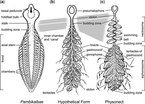
These chambers are big. Bigger than anything else so far found. Here's a reconstruction. Pambikalbae is clearly not a pennatulacean or cnidarian 'sea pen' like Charnodiscus. However, it does contain characters that suggest an evolutionary grade of organisation comparable to known cnidarians. The configuration and size of the chambers seems ideal to house symbiotic algae or bacteria . . . however, here's some wild speculation.
There is a group of cnidarians which do share similarities with Pambikalbae, and that's the physophorid Siphonophorida. Here's one below (photo credit)
Interesting isn't it. The chambers are especially similar. However, there is one thing wrong. The physophorid is upside down. The chambers actually point downwards. Why? Because physophorids float, they are not anchored to the sea floor like Pambikalbae.
But it's not as problematic getting from Pambikalbae to a physophorid, as it would appear.
(a) inverted Pambikalbae. (b) a hypothtical ancestral 'calycophore' siphonophore. (c) generalised modern physonect.
Just change a water-filled bulb as an holdfast, for an air filled bulb for a float. Simple folding inwards of the holdfast could produce the physonect and ancestral calycophore pneumatophore. A futher point of comparison is that the vane of Pambikalbae has three serial rows of chambers, as in calycophore and physonect siphonophores.
So a frondose form with wanderlust as ancestral to the modern siphonophore cnidarians? Maybe. But is could also be a derived hyrozoan, or a sister group to the early Chondrophorina. Whatever it is, it ain't no pennatulacean!
Jenkins, R.J.F. and Nedin, C. (2007) The provenance and palaeobiology of a new multi-vane, chambered frodose organism from the ediacaran (later Neoproterozoic) of South Australia. In P. Vickers-Rich, P. and Komarower, P. (eds) The Rise and Fall of the Ediacaran Biota. Geological Society Special Publication 286, 195-222. doi: 10.1144/SP286.15
Ignore the cute baby buffalo. See that unprepossessing roadside quarry with the people sitting on a pile of rubble (click to enlarge)? Well that rubble is the type section for the Chengjiang fauna.
Yeah! Not so unprepossessing now, is it?!
The Chengjiang fauna occurs in the Maotianshan Shale, a member of the Lower Cambrian Chiungchussu formation. They are found about 5 km northwest of the Fuxian Lake, and 6 km northeast of Chengjiang, Yunnan, China.
The age of the fauna is between 525-520 million years old, significantly older than the Burgess Shale (at 505 million years ago), and the Emu Bay Shale (approx 515 million years old).
The Chengjiang fauna rivals that of the Burgess Shale in preservation and diversity. Although not in difficulty to get to. The Burgess Shale is a 3 hour hike. Here, you can drive right to the outcrop!
In this shot (click to enlarge) you can see the quarry from photo one in the middle distance. There is a bus parked next to it with some people in the road. The hill to the right is Maotian Mount (hence Maotianshan Shale)
In this photo (click to enlarge), I am right at the back in short sleeves and a broad-brimmed brown hat (I'm in the same position in the first photo). I'm there partly because it's in the shade, and partly because there were some nice fossils there (all went to Nanjing University for study).
The depositional environment was delta front prograding eastwards into an open sea. Most of the fossil layers appear to be episodic events onto the marine muds in front of the delta, possible storm induced deposition of clays and fine sand. There's not much evidence of transport, so most organisms were locals and were buried by a series of turbidity flows.
This is a smaller quarry close by (click to enlarge). The dark colour is the fresh colour of the Maotianshan Shale. It is black when fresh, but rapidly oxidises to a tan colour on exposure to air. This is near the top of the Maotianshan Shale. The pick axe handle in the middle of the shot is marking the topmost Maotianshan Shales. The very top of the handle is resting against the first influx of sands, which coarsen upward until they are topped by a large lenticular sandstone at the top of the sequence above my head. This represents the prograding delta as it moves out over the muds of the Maotianshan Shale - just like the sands of the Mississippi Delta are prograding out into the Gulf of Mexico.
Look, I'm sorry about this. It's quite embarrassing, I know. But in my defense, I have blurred the naked Dickinsonia at the top right of the slab so as not to offend any sensibilities.
Anyhow, on to the real subject of this Palaeoporn, the structure with the highly descriptive and emotive name - "Form D". Yeah . . . "Form D" . . . no, honestly!
The Form D is the . . . um . . . rather flaccid structure above the compass. Form D was the name given to a group of thick, long, trace fossils of unknown origin. While they are not common, it doesn't take much poking about to expose them.
Given the lack of structure it was hard to attribute a source for the traces (they are not ribbed for example) thus making it difficult to erect a reasonable phylogeny and nomenclature. So they got stuck with the name "Form D" - as they were the fourth type of trace structures to be catagorised. Recently, they are thought to be, not trace fossils, but body parts. They appear to be association with the form Phyllozoon.
Phyllozoon Hanseni (yellow arrows) and Form D (blue arrows) from the Ediacaran of Canada.
Phyllozoon is a more-or-less bisymmetrical leaf-shaped structure composed of a series of 'tubes' which meet at a central zigzag suture. There seems to have been only one layer of tubes forming the 'vanes' of the frond, with a stiff outer covering. Numerous individuals are commonly scattered over bedding planes, sometimes overlapping. The fronds have obvious polarity (top and bottom) and their common association with Form D is too frequent to be chance. It is possible that Form D is actually the stolon to which the Phyllozoon fronds were attached. They may well have looked something like this below, except Phyllozoon probably wasn't green.
The alga Caulerpa taxifolia doing a passable imitation of Phyllozoon. Photo credit.
I suppose we should be grateful that Phyllozoon wasn't called Phallozoon . . .
You never forget your first. The time. The place. The moment.
The windswept coast. The bed that refused to cooperate. A cleavage that simply wouldn’t quit. But then suddenly, everything is revealed. The curves! So different. Stood out in the crowd despite being petite. I couldn’t fail to see, even at a distance of 4 feet.
No, you never forget your first. First meraspid that is.
This fellow is a personal favourite of mine. It’s a meraspid (or juvenile) Estaingia bilobata It was the first meraspid found at from the Emu Bay Shale. It’s a stage 10 meraspid. Thanks to it, and other merasipds found in the Emu Bay Shale, we can work out the ontogeny or growth pattern of Estaingia, and as an added bonus allows us to work out some evolutionary relationships (but that’s for another post).
This Palaeoporn is all about ontogeny amongst the trilobites, more specifically Estaingia.
One of the great things about trilobites is that, to grow, they have to moult the hard exoskeleton. This means that one trilobite can leave behind a complete growth history in moults. Since this is unlikely to happen for one, many trilobites of the same species leaving moults behind allows us to build up a picture of the growth pattern in a way that we simply can't do for moult-challenged organisms.
Meraspids are what trilobite larvae grow up to be before they grow up to be adult trilobites. Trilobite larvae, or protaspids look like just a head. Suddenly things get serious and the protaspid gets a tail. Or at least the beginnings of one. A furrow appears towards the back of the protaspid, which will separate the tail or pygidium from the head or cephalon. Once the furrow is complete, the trilobite is considered to have moved into the meraspid stage - meraspid 0. 0 because it has no thorax segments yet. Just a head and a tail.
Once in the meraspid stage, the trilobite begins to add segments to the thorax, usually one per moult, but in some species this can be more. Each meraspid degree or stage is represented by the addition of another thorasic segment and is numbered according to the number of segments.
The specimen at right (Fig. A) is a Meraspid 1, the smallest meraspid so far found at Emu Bay.
The most striking thing (apart from it's size - the white scale bar represents 1 millimetre! Hence it's a bit blurry) is the spines. In very small forms spines become important. If meraspid 1 was swimming amongst the plankton, the spines would help with buoyancy, helping keep the meraspid afloat. If the meraspid was on the sea floor, then the spines would help keep the meraspid from sinking into the mud. We don't know if Estaingia meraspids were planktonic or scurried around on the sea floor. The spines may also provide some defense, but the get smaller as the meraspid grows so it doesn't appear that defense was a primary function. In meraspid 1 the one segment has a pair of large spines, as well as a pair of backward pointing spines on either side of the head. Meraspid 2 would have had two segments each with a pair of long spines.
As the meraspid grows, segments are added from the front of the tail. We can see this in meraspid 7 at right (Fig. B, scale bar 1 mm). This has 7 segments, but the two original segments with the spines can be seen at the front of the thorax, with the non-spiney segments, which have been added later, positioned behind these two. If the thorasic segments were added from the back of the head, then the first two segments with spines would be at the back of the thorax next to the tail. The large spines on the head are also still very visible.
In Meraspid 10 at right (Fig. C, scale bar 1 mm) things are starting to change. The first thing to notice is that the large spine on segment 1 is almost gone. There is a much reduced spine there. Also the spines have become more curved. They are still long though - still the length of the meraspid. The head is very well preserved and provides a wealth of information on the way the various elements of the head change with growth. The rounded shape and the spines are very distinctive. Nothing else in the fauna is like that. So, even though it's only a few millimetres across, it immediately caught my eye.
Finally we have meraspid 12 (Fig. D, scale bar 1 mm). We are almost at the Holaspid stage or mature stage, of 13 segments, and the meraspid is now similar to the final adult shape. Note that the second thorasic spines are much reduced, and the spines on the head are also beginning to reduce in length. The thorasic spines will completely disappear in the adult, and the spines on the head will be reduced, but still present.
And this at right is a fully mature Estaingia. You can see that the thorasic spines have gone, and the spines on the head are much reduced.
Below is a diagrammatic representation of the ontogeny of Estaingia featuring the four meraspids figured above.
It gives you a better idea about what changes are occurring as the trilobite matures. Features to look out for are the relative width and length of the head and the glabella or central bulb region of the head. This is important because these relative values can be plotted and, as they remain at a constant ratio can help confirm the identification of the meraspids with the holaspid form. Like so:
This is a plot of the length of the longest part of the head (cranidium) against the width from eye to eye, and the longest part of the head against the longest part of the glabella - the central bulb area of the head.
As you can see the meraspid measurements (in red) line up along the same trend line as the adults (in blue) giving us a high degree of confidence that the meraspids are of Estaingia. Also notice that the measurements are clustered - i.e. not evenly distributed. This represents the moulting habit of the trilobites. As growth can only occur once the trilobite has moulted the pattern of growth is stepped. Unlike a constantly growing form, where the plot would be more even.
Using measurements of growth patterns can be very useful because with some trilobites, varying the time spent in particular growth patterns as can dramatically alter the end result - or what we call evolution. But that's for another post.
You may want to check out Palaeoporn 14 and 15 before reading this as Palaeoporn 16 follows on from them. That's ok. We'll just talk amongst ourselves until you get back.
So . . . where are you from? . . . Fort Saint John, Canada? Really? Good ol' Highway 97. . . . umm, come here often? . . .
Oh, ok you're back. Right. First thing, you need to see this video of a giant spider crab moulting (hat tip PZ)
Once you've seen it and read PZ's piece on moulting, or ecdysis. The linked video of a spider crab moulting in PZ's piece is the one featured here. Very instructive, if not a little creepy. But remember that trilobites - being proper arthropods - exited the old body exoskeleton old school i.e. head first, not arse first like a common chelicerate.
Ok, now we have an idea of what ecdysis is, this Palaeoporn is an example of what happens when things go wrong . . . very wrong.
The photo at the top is of the trilobite Redlichia takooensis, or rather what's left of one after a particularly difficult mould. Just to remind you, at right is what a Redlichia takooensis should look like. R takooensis is a Lower Cambrian trilobite with a 15 segment thorax and small pygidium or tail. A major characteristic of R takooensis are the large spines, on either side of the head, at the back of the head in the centre, and two large spines on the axial rings of segments 6 and 11 (counting back from the head) (What? Axial ring. The middle part of the thorax. What? Thorax. The middle part of the animal between the head and the back end (pygidium). What? Yes the pygidium is very small, it's a micropygous trilobite. What? No. The division head-thorax-pygidium is not how the trilobite got it's name. The "three lobes" refers to the three zones of the thorax, the plura on the left, the axial ring, and the plura on the right).
There are probably spines on each axial ring, but 6 and 11 have the biggest. The spine coming off the rear of the trilobite, seen in the photo (above right), is from segment 11 - so the spines were pretty long.
To give an idea of what R. takooensis looked like I'm going to cheat an use another trilobite to approximate Redlichia.
Left side view of Paradoxides masquerading as Redlichia (it's got three extra segments - no, don't count them!). Thick line represents facial suture. (adapted from Whittington 1990)
Notice the spines. These will be very important in what happens in extreme ecdysis. In life, the spines would have not sat flat along the thorax, but would have been mobile and could be moved in an arc, from flat along the body (as above and in complete trilobite photo above right), through some acute angle (as seen in the ParaRedlichia diagram above).
What you don't see is the series on notches on the back margin of the first 5 segments of the thorax. These would have acted to lock the first 6 segments in place making this portion of the thorax act as a rigid body. That's also important.
What's also important is that the trilobite could bend. It could flex into an arc - though not into a ball like later trilobites (an important feature for Anomalocaris as discussed previously). Also the maximum flexure occured around segment 5.
OK, ecdysis in Redlichia. Normally Redlichia would have operated in a similar way to Paradoxides shown below.
Maneuvers during exuviation of Paradoxides 2. Left side view, thick line on head represents the suture line. 3. Flexure of body. 4. Front view showing open sutures and animal beginning to emerge (stippled). 5. Side view of animal beginning to emerge. (Whittington 1990).
The trilobite arched upward, planted the front of the head in the sediment and pushed using the back end as a brace. This put pressure on the head and the sutures around it. These part, with the cheeks on either side parting from the central area of the head, and the front part of the head separating and lifting upwards. This allowed the soft new body to emerge through the front of the head and 'head' off, leaving the cast of exoskeleton behind.
There is one difference (alright two). The spines. These could have been brought into play if the old exoskeleton didn't play ball, as shown below.
Here, the rear spine could have been used as a lever to apply more pressure on the head sutures, forcing them to separate and allow the trilobite to exit. How do we know this? well, a) it seems like a reasonable thing to do, and b) we have the specimen figured as Palaeoporn 16 (and others).
Let's remind ourselves of the specimen, 'cos its been a while.
What we have here is one disarticulated trilobite. There is a split in the thorax between segments 4 and 5, and another between segments 10 and 11. The thorax has been split into three pieces, comprising segments 1-4, 5-10, and 11-15 (plus pygidium).
Which brings us to a very important question. Where's the head?
The left free cheek is there, roughly where it should be at the front end, but it's inverted. It's inside out. Flipped over. The nice curved margin you can see is actually where it joined the eye. But the rest of the head?
Well, most is missing, but see the white and orange line running away from the free cheek and pointing to the 2 o'clock position? That's what's left of the head. It's the lower part of the head, basically a thin band of exoskeleton (called the doublure) which runs around the underside of the head and attached to both free cheeks and the front of the central portion of the head (the doublure is show in black at right). That central top portion of the head is gone, but the doublure is rammed into the sediment and twisted.
To give you some idea, here is a somewhat stylised diagram to illustrate.
What we have is one very sad and sorry trilobite. But, knowing what we know of ecdysis in Redlichia we can piece (Ha!) together what happened.
Knowing that the first 6 segments could lock together, and and the spine on segment 11 was used as a pivot point, simple mechanics tells us the the maximum forces exerted at or around segment 5, (where the maximum curve would be - the locking of segments 1-6 would re-enforce this), and around segment 11.
So, the trilobite arched up, rammed the front of the head into the sediment and braced with the spine on segment 11. Nothing happened.
Eventually the arching and the bracing built up so much pressure that something had to give. In very quick succession, the exoskeleton behind the head went (we know this can happen from Palaeoporn 15), as did the sutures on the head. But also the pressure acting on segment 5 became too much and the exoskeleton ruptured between segments 4 and 5. Also to pressure around segment 11, caused by bracing against the spine, causes the exoskeleton to rupture between segments 10 and 11.
The trilobite exited the broken exoskeleton, probably taking the middle portion of the head with it. That left the free cheeks separated from the central head exoskeleton, but still attached to the doublure, which was buried in the sediment. The left free cheek, still attached on its outer side, rotated outward to lie inside out, while the other is buried.
Tough moult!
But there's more. Here's another example. Similar story with similar results (sorry it's in black and white. I could say it's in black and white for the ambiance, but I just don't have a colour photo of it).
This time the thorax exoskeleton gave way behind segment 6 and behind segment 11. And this time we have the head. And both free cheeks, but they are inverted an displaced slightly. Also the doublure is present. it's the thick band right at the front of the head.
This is a classic example of a broken, incomplete, trilobite providing more information than a perfect specimen would. They provide information on trilobite activity and mechanical information on the exoskeleton.
They also provide a whole new definition of the term "growing pains"
Whittington, H.B (1990) Articulation and Exuviation in Cambrian Trilobites. Phil. Trans. R. Soc. Lond. B. Vol. 329 no. 1252. pp:27-46 doi:10.1098/rstb.1990.0147
These are Estangiabilobata trilobites from the Lower Cambrian Emu Bay Shale on Kangaroo Island, South Australia. Estangia is the most common fossil found in the Emu Bay Shale. However, these came from the outcrop of Emu Bay Shale at Emu Bay, and not from the more famous site further along the coast that contains exceptionally preserved fossils such as Anomalocaris and Myoscolex. (The Emu Bay Shale outcrops at two locations on Kangaroo Island)
These little critters show the difference between the two depositional sites. The site with exceptional preservation shows evidence - palaeontological (whole specimens, low diversity) sedimentological (fine grained sediments), and chemical (evidence of reducing environment-enriched trace elements) - of a low energy, low oxygen environment conducive to exceptional preservation.
On the other hand at Emu Bay the site shows evidence of a higher energy, higher oxygen depositional environment. This is because the sediments show more interbedded sands and silts (coarser grained therefore higher energy), oxidating environment-enriched trace elements, and the fossils do not show exceptional preservation, and are fragmented.
In the example above, the fossils assemblage comprises the heads of three Estangia trilobites (the lower one is both turned over and spun through 180 degrees). The heads are not complete. The sides of the head - the librigena (or the free cheeks) are missing. This shows that the heads represent molts.
Trilobites are arthropods and so have to molt the outer exoskeleton in order to grow. To do this, they have special lines of weakness in the exoskeleton called sutures. When the trilobite molts, these suture lines break apart, allowing the trilobite to leave the exoskeleton. These sutures are particularly obvious on the head where they run from the margin down to the eye, around the eye and then back out to either the side margin or the back margin.
In this case the suture lines are opisthoparian, as they run from the eye to the back of the head rather than out to the side They run along the eye so that the eyes will be the first thing to break out of the old exoskeleton - allowing the trilobite to keep its vision while the molting process continues. This results in the librigena breaking away from the head. Often the molting process breaks the attachment between the head and the rest of the body, resulting in the head becoming detached from the rest of the body.
Since these fossils comprise the cranidium only (head minus the librigena), this indicates that they are molts and that they have been sorted by currents that have separated them from the body and librigena.
Compare this with another Emu Bay Shale Estangia, this time from the site of exceptional preservation (right).
Here the head and body is present. However, it is still a molt because the librigena have been freed from the head. In this specimen, the right librigena (outlined) is still associated with the body but has moved some distance away, and is both turned over and spun through 180 degrees so the the spine (that normally points backwards) now points forwards.
So the three in the top image represent a disarticulated random grouping, and have not been caught in flagrante delicto. So move along . . . nothing to see here . ..
Look up there! Is it a brachiopod? Is it an annelid? Is it a mollusc?
Umm . . . actually, its a bit of each really.
This is a wiwaxiid from the Emu Bay Shale. Unfortunately undescribed, but closely linked to Wiwaxia and the halkieriids. It would be one of the oldest examples of a wiwaxiid, as the others are from the Middle Cambrian Burgess Shale and comparable deposits.
The beast was bilaterally symmetrical, oval in shape, and covered with short scales - called sclerites. Also present were a number of rows of larger spines which protruded upwards in a probable defensive array (see reconstruction at right)
Wiwaxiids had a flat foot-like underside. Little is known of the internal anatomy.
Wiwaxiids have been classified as molluscs, annelids and stem group annelids (a group closely related to annelids). The spines have been compared to the eltrya, or scales, of polychaete worms, and even bristles of molluscs, annelids and brachiopods, and halkieriids, of course, have little brachiopod shell caps!
One feature found in wiwaxiids is a radula-like feeding bar. So I looked to see if my specimens had a radula-like feeding bar. I've switched to black and white photos for higher resolution. A bar structure, formed of calcium phosphate, was found towards the front of the specimen. After preparing out (lower image) half the bar remained and the other half (the distal, or outer, part of the bar is removed, leaving a mold of the lower surface of the bar. in this mold can be seen several depressions along the 'upper' margin, represented by shadow (the light is coming from the top right of the image). These are 'teeth' which would have protruded from the lower margin of the bar (the bar is approx. 5 mm long.)
So, spines, radula-like feeding bar, seems like a Lower Cambrian wiwaxiid!
Naraoia (above, scale = 1 cm) has featured here before (what? . . . I know it's not monochromatic, just wait), but this time I’m going to show what a little black and white photography can do (see!).
First of all, this (right) is what Naraoia looked like in the flesh. It had two ‘shields’ an anterior, or head shield, and a posterior shield. These shields overlapped, so that the posterior end of the head shield rested over the anterior edge of the posterior shield.
Underneath the shields, the body looked very much like that of trilobites, to whom they are closely related. In other words they had a many segmented body (including the head), and a pair of biramous appendages on most segments.
Fossils from the Lower Cambrian Emu Bay Shale are not exceptionally well preserved when it comes to fine structure. The fossils have been recrystallised into hulking great fibrous calcite crystals during a subsequent tectonic event. But, they do look good in colour, even the one above which is not a particularly good specimen.
However, occasionally you can get some detailed information, and then the best documentation is obtained using black and white photography, which is why this is still the preferred medium for journals, especially where structure is indistinct and a bugger to document!
All the photography here was done the old fashioned way, before that new-fangled digitally enhancement, with honest to goodness film, and dark rooms, and the inhalation of toxic chemicals during development and printing, Didn't effect me though, but I digress . . . err, who am I again?
Basically light and shade can show up very fine structures that colour cannot. Maybe because we can interpret subtle changes in greyscale as meaningful structures, whereas this is more difficult in colour, I don’t know. But by varying the intensity and the angle of the light, and by using high magnification and very slow film (in this case Ilford 100 and Ilford Pan F Plus) structures can be seen that are either absent of very difficult to see in colour photos.
Below I’ll show a couple of examples of what I mean.
Here is a black and white image of another specimen of Naraoia (click to enlarge). This time you can see some structure. The image on the right has been augmented to show them. Other than that the image has not been manipulated, and the structures are seen because the angle of the light hitting the specimen is very low.
A number of structures in the head can be seen, including the antennae, legs and a remnant of the hypostome, which is a rigid plate that sat directly in front of the mouth. A very thick doublure is also present. This is where the posterior shield has folded back on itself around the margin. This strengthens the shield.
The area where the two shields overlap can also be seen. Now this image is a scan of a negative that has been converted to a JPEG file and then uploaded, so some detail has been lost, but they are there . . . trust me, I’m a doctor!
Still not convinced? OK, how about this:
This image is a magnification of the last (click to magnify further), but with a slight change in the light intensity, and the angle. Other than that, and the augmentation of the right hand image, there has been no manipulation. The field of view is approximately 1 cm by 1.5 cms. Despite the fact that the magnification resulted in a very small depth of field, a number of structures are visible, in fact more so that in the last image.
There are at least 6 head appendages behind the antennae (at a push), and the hypostome is visible. Also, the margins of the anterior and posterior shields are there.
There is also a lot of wrinkling. This is because, in life, the critter was quite domed, and that has been flattened out leaving the mass of wrinkles at the front of the head. Also the left hand margin of the posterior shield is twisted near its anterior margin, and a number of major wrinkles have formed on the posterior shield in response to this twist.
If you can't document your structures, you can't publish them. Which is fine if you are dealing with Burgess Shale levels of preservation. However, the rest of us have to rely of long hours in the photography suite playing with magnification, light intensity, and lighting angles. That's before you get to the developing and printing phase.