The answer is that, in monotremes, the duct is inside the orbit, just like placentals.
 The skull of an Echidna (above) shows the duct to be inside the orbit. In an actual specimen, the duct is placed in the bone surface sloping into the orbit (not clear on a two-dimensional photo). In a marsupial the duct would be clearly visible outside the orbit.
So there you go, only marsupials have the duct visible outside the orbit.
So the question now is, how do you tell a placental anteater skull from an Echidna skull?
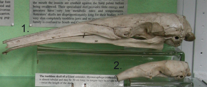
The answer is that the Echidna is far more bird-like than the anteater, and the cranium tends to slope sharply downward at the front in the Echidna, but tends to be much flatter in the anteater (see below).

Photo credit
Echidna - University of Washington
Anteater - Natural History Museum, London
This is really interesting, considering that some folks in the past have considered monotremes to be potentially far removed from the rest of crown Mammalia and possible sister taxa of Multituberculata. I guess that these external nasolacrimal ducts could be derived for marsupials, correct?
ReplyDeleteIt looks like it is on the outside on tamanduas
ReplyDeleteand giant anteaters but maybe inside on pygmys.
hyyp://www.azdrybones.com/images/LesserAnteater.jpg
Hope that works as copy paste seems to be disable but tried to type it right.